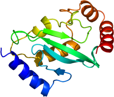
E2-EPF5: Human Ubiquitin Carrier Protein
PDB:1ZDN
Revision
Revision Type:created
Revised by:created
Revision Date:created
Entry Clone Accession:
Entry Clone Source:MGC
SGC Clone Accession:ubc52.001.156; plate SDC013D:10
Tag:N-terminal His-tag with integrated thrombin-cleavage site MGSSHHHHHHSSGLVPRGS.
Host:E.coli BL21 (DE3)
Construct
Prelude:
Sequence:gsMNSNVENLPPHIIRLVYKEVTTLTADPPDGIKVFPNEEDLTDLQVTIEGPEGTPYAGGLFRMKLLLGKDFPASPPKGYFLTKIFHPNVGANGEICVNVLKRDWTAELGIRHVLLTIKCLLIHPNPESALNEEAGRLLLENYEEYAARARLLTEIHG
Vector:p28-LIC-Thrombin
Growth
Medium:TB
Antibiotics:
Procedure:Bacterial cells, E. coli BL21 (DE3) transformed with the target protein cDNA inserted into p28-LIC-Thrombin vector and stored as glycerol stubs at -80°C, are grown in 3 mL LB medium (Sigma L7658) supplemented with 50 µg/mL kanamycin monosulfate (BioShop Canada KAN 201) for 8 h at 37°C in a shaking incubator. Two mL of this culture is used to inoculate 100 mL of kanamycin/LB in a baffled flask, and cell growth continues overnight at 37°C with shaking. Next morning, the resulting preculture is used for inoculation of 2 L TB medium (Sigma T0918) containing 0.15% glycerol (EMD GX0185-5), 50 µg/mL kanamycin and 200 µl Antifoam 204 (Sigma A-8311). Cells are grown at 37°C with vigorous aeration to an OD600 of 4-6. The cell culture is cooled to 15°C, induced with 50-100 µM isopropyl-thio-D-galactopyranoside (BioShop Canada IPT 001), and incubation at this temperature continues overnight with constant aeration. The culture is divided between two plastic bags (Beckman Coulter 369256) and centrifuged for 15 min at 7,000 rpm in a JLA 8.1000 rotor using Avanti J-20 XPI centrifuge (Beckman Coulter). The supernatant is discarded and cell pellets in the bags are frozen and stored at -80°C.
Purification
Procedure
Immobilized-metal Affinity Chromatography: An aliquot (20 µl) of clarified supernatant is reserved for later analysis by SDS-PAGE. To the rest of it, 6 mL of 50% suspension of TALON metal-affinity resin (BD Bioscience) in the lysis buffer is added (3 mL suspension per each 50-mL tube with the supernatant). The tubes are incubated for 1 h in refrigerator with constant stirring, centrifuged at 4°C for 2 min at 1,000 rpm in SX4750 rotor using Allegra X-12R centrifuge (Beckman Coulter) and the supernatant is discarded. The affinity resin pellets are suspended in 10 mL lysis buffer and transferred into an Econo-Column (Bio-Rad 732-1010). The buffer is allowed to drain and discarded. The settled gel is washed with 15 mL lysis buffer containing 10 mM imidazol (washing buffer) followed by 15 mL washing buffer containing 0.05% Tween 20 (Sigma P7949) and 30 mL washing buffer; the washings are discarded. Elution is achieved using lysis buffer containing 200 mM imidazol. First 0.5 mL eluate is discarded and two 5-mL fractions are collected after that in tubes containing 50 µl 1M DTT (final DTT concentration is 10 mM). Protein concentration in these fractions is determined using Coomassie Plus Protein Assay reagent (Pierce 1856210) and bovine serum albumin (Pierce 23209) as a protein standard for calibration. The recombinant protein is usually eluted in the first 5-mL fraction and it is used directly at the next purification step. Some His-tagged proteins spread between the two fractions. In this case, the fractions are combined and concentrated down to approx. 5-mL volume by ultrafiltration (Amicon Ultra-15 10,000 MWCO, UFC901024 or 5,000 MWCO, UFC900524, as appropriate, Millipore).
Size-exclusion Chromatography: This step is performed using an ÄKTA Purifier or an ÄKTA Express system (GE Healthcare). The protein sample is loaded onto an XK16x65 column packed with HighLoad Superdex 200 (GE Healthcare) and equilibrated with 20 mM Tris-HCl, pH 8.0, 500 mM NaCl, 5% glycerol, 10 mM dithiothreitol (BioShop Canada, DTT 001). Elution is performed with the same buffer at a flow-rate of 3 mL/min in the Peak Fractionation mode: {Slope; min. peak width 0.833 min; level 0.000 mAU; peak start slope 10.000 AU/min; peak end slope 20.000 AU/min}. Note: If target protein has OD280 (0.1%, 1 cm) lower than 0.2, monitor the eluate absorbance at 215 ηm and use either time/volume-based or manual fraction collection. Fractions corresponding to major peak on the chromatogram are combined and analyzed by SDS-PAGE and LC/MS. Protein concentration is determined by measuring UV absorbance of the combined fractions at 280 ηm. Purified protein is concentrated using an ultrafiltration concentrator as above to a final concentration of approx. 50 mg/mL for further crystallographic screening (preceded by a PC test, Hampton Research, in order to determine optimal protein concentration) and biophysical characterization. The concentrated protein solution is divided into 100-µl aliquots that are frozen by immersion in liquid nitrogen and stored at -80ºC.
His-tag Removal: The purified protein, 10-20 mg, is diluted in 50 mM Tris-HCl, pH 7.5, 140 mM NaCl to a final volume of 4 mL and thrombin (Sigma T9681) is added (1 unit per mg target protein). The reaction mixture is incubated for 2 h at 21-23°C, and the protein is re-purified using size-exclusion chromatography as above.
Extraction
Procedure
Frozen cell pellets in the plastic bags are thawed and placed on ice. Each cell pellet is resuspended in 30 mL lysis buffer [50 mM Tris (VWR EM-9210)-HCl (EMD ACS393-42), pH 8.0, 250 mM NaCl (VWR EM-SX0420-1), 1mM ß-mercaptoethanol (Fluka 63689), 5% glycerol, 0.1mM phenylmethanesulfonyl fluoride (Sigma P7626), 1 mM magnesium chloride (Fluka 63063), 100 ?M zinc chloride (Fluka 96468) and 2 mM imidazol (BioShop Canada IMD508)] and homogenized with a 30-second burst of Ultra-Turrax T8 homogenizer (IKA Works) at maximal setting. Cell lysis is accomplished by passing the cell suspension through Microfluidizer Processor M-110EH (Microfluidics) at 18,000-20,000 psi setting with ice cooling. Lyzed cells are centrifuged at 4°C for 20 minutes at 23,000 rpm in a JA25.50 rotor using Avanti J-20 XPI centrifuge (Beckman Coulter). The supernatant is decanted into two sterile 50-mL polypropylene conical tubes (Becton Dickinson Labware 0420705), and the pellet is discarded.
Concentration:20 mg/mL
Ligand
MassSpec:
Crystallization:Purified human E2-EPF5 was crystallized using the hanging drop vapor diffusion method. One µL of the diluted protein solution (10 mg/mL) was mixed with 1 µL of the reservoir solution consisting of 3.25M Na formate, 0.1M bisTris, pH 7.0 at room temperature (293K).
NMR Spectroscopy:
Data Collection:
Data Processing: