PPIL2
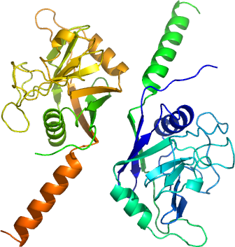
PDB:1ZKC
Revision
Revision Type:created
Revised by:created
Revision Date:created
Entry Clone Accession:GI:22547214
Entry Clone Source:MGC
SGC Clone Accession:ppi65.269.457:A2; plate SDC023 A2
Tag:N-terminal histag with integrated thrombin cleavage site: mgsshhhhhhssglvpr*gs
Host:BL21(DE3)
Construct
Prelude:Sequence:mgsshhhhhhssglvprgsGYVRLHTNKGDLNLELHCDLTPKTCENFIRLCKKHYYDGTIFHRSIRNFVIQGGDPTGTGTGGESYWGKPFKDEFRPNLSHTGRGILSMANSGPNSNRSQFFITFRSCAYLDKKHTIFGRVVGGFDVLTAMENVESDPKTDRPKEEIRIDATTVFVDPYEEADAQIAQERKTQLKIAP
Vector:p28a-LIC
Growth
Medium:TB
Antibiotics:Procedure:Using the SGC's
LEX bubbling system, PPIL2 was expressed in E. coli BL21 (DE3) grown in Terrific Broth (TB) in the presence of 50 microG/mL of kanamycin at 37degC to an OD
600 of 7.5. Cells were then induced by isopropyl-1-thio-D-galactopyranoside (IPTG), final concentration 0.05 mM, and incubated overnight at 15ºC. The culture was centrifuged and the cell pellets were collected and stored at -80degC.
Purification
ProcedureIMAC purification: 4 microL of clarified supernatant is reserved for later analysis by SDS-PAGE. The rest of the clarified supernatant is then diluted 1:2 in lysis buffer, and loaded at approximately 1mL/min by gravity onto 5 mL of Ni-NTA resin (Qiagen 30450). 5 column volumes of lysis buffer are used to wash the column at approximately 3 mL/min, followed by 5 column volumes of low imidazole buffer at approximately 3 mL/min. A 4 µL sample of the low imidazole wash is saved for later analysis by SDS-PAGE. Samples are eluted from the Ni-NTA resin by exposure to 10 mL elution buffer at 1mL/min flow rate. A 10 µL sample of the eluate is saved for SDS-PAGE analysis. 10 microL of each eluate is saved for measurement of protein concentration using Bradford reagent (BioRad 500-0202).
Size exclusion chromatography: An XK 16x65 column (part numbers 18-1031-47 and 18-6488-01, GE Healthcare) packed with HighLoad Superdex 200 resin (10-1043-04, GE Healthcare) is pre-equilibrated with gel filtration buffer for 1.5 column volumes using an AKTAxpress (18-6645-05, GE Healthcare) at a flow rate of 3 mL/min. 5 mL of sample is loaded onto the column at 1.5 mL/min, and 2 mL fractions are collected into 96-well plates (VWR 40002-012) using peak fractionation protocols with the following parameters: (Slope; min. peak width 0.833 min; level 0.000 mAU; peak start slope 10.000 AU/min; peak end slope 20.000 AU/min). Peak fractions are analyzed for purity using SDS-PAGE or visual analysis of the chromatogram and pooled.
Concentration: Purified proteins are concentrated using either 4 mL or 15 mL concentrators with an appropriate molecular weight cut-off (Amicon Ultra-15 10,000 MWCO, UFC901024 or 5,000 MWCO, UFC900524, as appropriate, Millipore) to a final concentration of 20 mg/mL for crystallographic screening or other biophysical studies.
Extraction
ProcedureFrozen cell pellets contained in bags (Beckman 369256) obtained from 2L liters of culture are thawed by soaking in warm water for 5 minutes. Each cell pellet is resuspended in 20 mL lysis buffer and 1mL Sigma general protease inhibitor (Sigma P2714-1BTL, resuspended according to manufacturerÂs instructions) and then homogenized using an Ultra-Turrax T8 homogenizer (IKA Works) at maximal setting for 30-60 seconds per pellet. Cell lysis is accomplished by sonication (Virtis408912, Virsonic) on ice: the sonication protocol is 10 sec pulse at half-maximal frequency (5.0), 10 second rest, for 6 minutes total sonication time per pellet. Lysed cells are placed into centrifuge tubes (363647, Beckman Coulter) and centrifuged in a JA25.50 rotor in an Avanti J-20 XPI centrifuge (Beckman Coulter) for 20 minutes at 69,673 x g. The supernatant is decanted into a beaker, and the insoluble pellet discarded.
Concentration:LigandMassSpec:Crystallization:Purified PPIL2 was crystallized using the sitting drop vapor diffusion method. Diffracting crystals leading to the structure grew when the protein was mixed at 20 mg/mL with the reservoir solution (containing 0.8M Potassium Sodium Tartrate tetrahydrate, 0.1M Hepes pH 7.5) in a 1:1 volume ratio.
NMR Spectroscopy:Data Collection:Data Processing: