Entry Clone Source: Ordered-synthetic |
Entry Clone Accession: n/a |
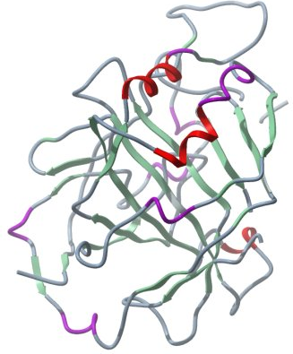
SGC Construct ID: CA8A-c010 |
GenBank GI number: gi|22027500 |
Vector: pNIC28-Bsa4. Details [PDF ]; Sequence [ FASTA ] or [ GenBank ] |
Tags and additions: Tag sequence: N-terminal His-tag with a TEV protease cleavage site: mhhhhhhssgvdlgtenlyfq(*)sm |
Tag removed: yes |
Expressed sequence (tag sequence in lowercase):
mhhhhhhssgvdlgtenlyfqsmADLSFI
EDTVAFPEKEEDEEEEEEGVEWGYEEGVE
WGLVFPDANGEYQSPINLNSREARYDPSL
LDVRLSPNYVVCRDCEVTNDGHTIQVILK
SKSVLSGGPLPQGHEFELYEVRFHWGREN
QRGSEHTVNFKAFPMELHLIHWNSTLFGS
IDEAVGKPHGIAIIALFVQIGKEHVGLKA
VTEILQDIQYKGKSKTIPCFNPNTLLPDP
LLRDYWVYEGSLTIPPCSEGVTWILFRYP
LTISQLQIEEFRRLRTHVKGAELVEGCDG
ILGDNFRPTQPLSDRVIRAAFQ |
Host: BL21(DE3)-R3-pRARE2 |
Expression protocol: Competent BL-21 (DE3)-R3-pRARE2 phage resistant cells were transformed with 6 µl of the plasmid DNA and plated out onto LB plate plus 50 µg/ml kanamycin and 35 µg/ml chloramphenicol. The next day colonies were picked into fresh deep well blocks containing 1 ml TB + 50 µg/ml kanamycin and 35 µg/ml chloramphenicol which were grown overnight and glycerol stocks were prepared by adding 333 µl of 60 % glycerol to 1 ml of cell suspension, which were stored at -80°C to be used for future scale up preparations. The glycerol stock was used to inoculate 10 ml of TB (terrific Broth) supplemented with 50 µg/ml kanamycin and 35 µg/ml chloramphenicol . This starter culture was grown overnight at 37°C and used to inoculate a 1 liter culture in the same medium. The culture was grown at 37°C until the OD600 reached ~3.0. After that the temperature was lowered to 18°C. Protein production was induced with 0.1 mM IPTG and recombinant CA8Awas expressed at that temperature over night. The next day cells were harvested by centrifugation at 4000 rpm for 20 minutes then the pellets were scraped out and transferred to 50-ml Falcon tubes and frozen at -80°C. |
Cell extraction: 2x Lysis buffer: 100 mM K-phosphate, pH 7.5, 1M NaCl, 1 mM TCEP, 1x Protease Inhibitors Cocktail Set VII (Calbiochem, 1/1000 dilution), and 15 units/ml Benzonase. Lysis buffer: 50 mM K-phosphate, pH 7.5, 0.5M NaCl, 1 mM TCEP. |
Procedure: Frozen cell pellets (g) were thawed briefly in a bath of warm water (37°C) then transferred to ice. One volume (i.e. 1 ml for every gram of cells) of 2x lysis buffer was added, followed by 1x lysis buffer to a total volume of 150 ml. The cells were resuspended by agitating and disrupted by high pressure homogenization (30 kpsi). Nucleic acids and cell debris were removed by adding 0.15% PEI (polyethyleneimine) from a 5% (w/v, pH 7.5) stock, stirring for 30 minutes, then centrifugation for 1 hour at 16,000 x g. The supernatant was then further clarified by filtration (Acrodisc filters, 0.2 mm). |
Column 1: Ni-affinity, HisTrap Crude FF, 5 ml (GE Healthcare) |
Buffers: Affinity buffer: 50 mM K-phosphate, pH 7.5, 500 mM NaCl, 10 mM imidazole, 0.5 mM TCEP; Wash buffer: 50 mM K-phosphate, pH 7.5, 500 mM NaCl, 30 mM imidazole, 0.5 mM TCEP; Elution buffer: 50 mM K-phosphate, pH 7.5, 500 mM NaCl, 300 mM imidazole, 0.5 mM TCEP. |
Procedure: The cell extract was loaded on the column at 4 ml/minute on an AKTA-express system (GE Healthcare). The column was washed with 10 volumes of lysis buffer, 10 volumes of wash buffer, and then eluted with elution buffer at 4 ml/min. The eluted peak of A280 was automatically collected. |
Column 2: Gel filtration, Hiload 16/60 Superdex S75 prep grade, 120 ml (GE Healthcare) |
GF buffer: 10 mM HEPES, pH 7.5, 500 mM NaCl, 5% Glycerol, 0.5 mM TCEP, 4 mM MgCl2. |
Procedure: The eluted fraction from the Ni-affinity Histrap column was loaded on the gel filtration column in GF buffer at 0.80 ml/min. Eluted proteins were collected in 2-ml fractions and analyzed on SDS-PAGE. |
Mass spectrometry characterization: ESI-MS revealed that the protein had a mass of 33060.3242 Da (expected mass: 33060). |
Compound: none. |
Protein concentration: Buffer was changed to 10 mM HEPES, 150 mM NaCl, 5% glycerol. The protein was concentrated to 10.5 mg/ml using a centricon with a 10kDa cut off. The protein concentration was determined spectrophotometrically using e280= 46410. |
Crystallization: Crystals were grown at 4°C by vapour diffusion in sitting drops mixing 100 nL protein (10.5 mg/mL) and 200 nL well solution containing 10% (w/v) PEG 6000, 0.3 M NH4Cl pH 6.3 and 10% (v/v) ethylene glycol. Crystals were cryo-protected using 20% (v/v) ethylene glycol and flash cooled in liquid nitrogen. |
Data Collection: Resolution :1.6Å; X-ray source: SLS beam 10XSA |