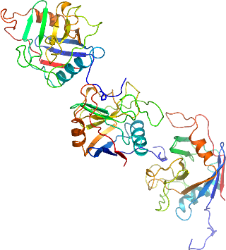
PPWD1
PDB:2A2N
Revision
Revision Type:created
Revised by:created
Revision Date:created
Entry Clone Accession:gi:24308048
Entry Clone Source:MGC
SGC Clone Accession:ppi60.461.646:E3; plate SDC022 E3
Tag:N-terminal His-tag with integrated thrombin-cleavage site MGSSHHHHHHSSGLVPRGS.
Host:E.coliBL21 (DE3)
Construct
Prelude:Sequence:gsPSKEEVMAATQAEGPKRVSDSAIIHTSMGDIHTKLFPVECPKTVENFCVHSRNGYYNGHTFHRIIKGFMIQTGDPTGTGMGGESIWGGEFEDEFHSTLRHDRPYTLSMANAGSNTNGSQFFITVVPTPWLDNKHTVFGRVTKGMEVVQRISNVKVNPKTDKPYEDVSIINITVK
Vector:p28a-LIC
Growth
Medium:TB
Antibiotics:Procedure:PPWD1 was expressed in E. coli BL21 (DE3) grown in Terrific Broth (TB) in the presence of 50 µg/ml of kanamycin at 37ºC to an OD600 of 7.5. Cells were then induced by isopropyl-1-thio-D-galactopyranoside (IPTG), final concentration 0.05 mM, and incubated overnight at 15ºC. The culture was centrifuged and the cell pellets were collected and stored at -80ºC.
Purification
ProcedureIMAC purification: 4 microL of clarified supernatant is reserved for later analysis by SDS-PAGE. The rest of the clarified supernatant is then diluted 1:2 in lysis buffer, and loaded at approximately 1mL/min by gravity onto 5 mL of Ni-NTA resin (Qiagen 30450). 5 column volumes of lysis buffer are used to wash the column at approximately 3 mL/min, followed by 5 column volumes of low imidazole buffer at approximately 3 mL/min. A 4 microL sample of the low imidazole wash is saved for later analysis by SDS-PAGE. Samples are eluted from the Ni-NTA resin by exposure to 10 mL elution buffer at 1mL/min flow rate. A 10 microL sample of the eluate is saved for SDS-PAGE analysis. 10 microL of each eluate is saved for measurement of protein concentration using Bradford reagent (BioRad 500-0202).
(His)6-tag cleavage: The 10 mL of Ni-NTA eluate from 2 is divided into 2x5 mL samples in 50 mL conical vials (352096, BD Biosciences). 5 mL of uncut sample is loaded onto gel filtration (see below); 1unit of thrombin (Sigma T9681) per milligram of protein is added to the other 5 mL sample. The conical vial is stored without shaking, overnight, at 4degC.
Size exclusion chromatography: An XK 16x65 column (part numbers 18-1031-47 and 18-6488-01, GE Healthcare) packed with HighLoad Superdex 200 resin (10-1043-04, GE Healthcare) is pre-equilibrated with gel filtration buffer for 1.5 column volumes using an AKTAxpress (18-6645-05, GE Healthcare) at a flow rate of 3 mL/min. 5 mL of sample is loaded onto the column at 1.5 mL/min, and 2mL fractions are collected into 96-well plates (VWR 40002-012) using peak fractionation protocols with the following parameters: (Slope; min. peak width 0.833 min; level 0.000 mAU; peak start slope 10.000 AU/min; peak end slope 20.000 AU/min). Peak fractions are analyzed for purity using SDS-PAGE or visual analysis of the chromatogram and pooled.
Concentration: Purified proteins are concentrated using either 4 mL or 15 mL concentrators with an appropriate molecular weight cut-off (Amicon Ultra-15 10,000 MWCO, UFC901024 or 5,000 MWCO, UFC900524, as appropriate, Millipore) to a final concentration of 20 mg/mL for crystallographic screening or other biophysical studies.
Extraction
ProcedureThe cell pellet was resuspended in lysis buffer + protease inhibitor (0.1 micromolar phenylmethyl sulfonyl fluoride, PMSF)and lysed using Microfluidizer. The lysate was cleared by centrifugation.
Concentration:LigandMassSpec:Crystallization:Purified PPWD1 was crystallized using the hanging drop vapor diffusion method. Crystals grew when the protein (12 mg/mL) was mixed with the reservoir solution in a 1:1 volume ratio, and the drop was equilibrated against a reservoir solution containing 1.7 M ammonium sulphate, 0.1 M sodium cacodylate 0. 2M sodium acetate at pH 5.7 in 293K temperature.
NMR Spectroscopy:Data Collection:Data Processing: