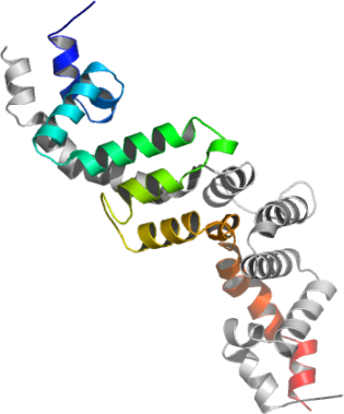
RGS7
PDB:2A72
Revision
Revision Type:created
Revised by:created
Revision Date:created
Entry Clone Accession:RGS 7A-s001
Entry Clone Source:MGC
SGC Clone Accession:Tag:N-terminal hexahistidine tag
Host:Bl-21(DE3)R3 phage resistant
Construct
Prelude:Sequence:SMKEPSQQRVKRWGFGMDEALKDPVGREQFLKFLESEFSSENLRFWLAVEDLKKRPIKEVPSRVQEIWQEFLAPGAPSAINLDSKSYDKTTHNVKEPGRYTFEDAQEHIYKLMKSDSYPRFIRSSAYQELLQAKKKSGNSMDRRTS
Vector:pNIC-Bsa4
Growth
Medium:Antibiotics:Procedure:TRANSFORMATION: 2 µL of each construct DNA was added to each well of a PCR plate on ice. 90 µL of competent BL21-(DE3)R3 bacteria were added to each well, with the plate remaining on ice. The plate was left on ice for a further 20 minutes. The heat-shock procedure was carried out by transferring the plate to a 42°C water bath for 45 seconds and then returning it to sit on ice for 2 minutes. 100 µL of SOC medium (pre-warmed to 42°C) was added to each well and the plate incubated at 37°C for 60 minutes. 150 µL of each culture was plated out onto LB-agar containing 33 µg/mL kanamycin in a 5.5 cm Petri dish. The plates were incubated at 37°C overnight.
PREPARATION OF GLYCEROL STOCKS: A number of colonies were used to innoculate 1mL of 2TY with 50 µg /mL kanamycin in a 96-well block. The block was placed in a shaker overnight at 37°C and 200 rpm. 40 µL of 70% glycerol was combined with 60 µL of bacterial culture in sterile 96-well microtitre plates. The plates were placed in the -80°C freezer.
EXPRESSION: 20 mL LB + 33 µg/mL kanamycin was inoculated using the glycerol stock and grown overnight. 16 mL of the sample was used to innoculate 2 litre TB medium + 33 µg/mL kanamycin . The culture was grown at 37° C for 2.5 hours before the incubator temperature was reduced to 25°C. The cultures were induced with 1 mM IPTG one hour after the temperature shift when the OD600 of the cells was approximately 0.9.
The cells were harvested by centrifugation 4 hours after induction. All the pellets were resuspended in Lysis Buffer and stored in a -80°C freezer.
Lysis/Binding Buffer: 50 mM Hepes pH 8.0, 500 mM NaCl, 5 % glycerol, 5mM imidazole pH 8.0, 0.5 mM TCEP
Purification
ProcedureWash Buffer: 50 mM Tris pH 8.0, 500 mM NaCl, 5 % Glycerol, 25 mM Imidazole pH 8.0, 0.5 mM TCEP; Elute Buffer: 50 mM Hepes pH 8.0, 500 mM NaCl, 5 % Glycerol, 250 mM Imidazole pH 8.0, 0.5 mM TCEP; GF Buffer: 50 mM Tris pH 8.5, 500 mM NaCl, 0.5 mM TCEP.
Procedure: The supernatent was filtered through a 1.2 micron and then a 0.2 micron syringe filter. A new 1 mL HisTrap column was used for IMAC. After washing with water the column was equilibrated with 5 column volumes of Binding Buffer at a flow rate of 0.8 mL/min. Binding of the sample to the resin was carried out by flowing the supernatant over the resin at a rate of 0.8 mL/min. Once bound the resin was washed with a minimum of 15 mLs binding buffer followed by a minimum of 20 mLs wash buffer. The application of 5 mLs of Elution Buffer eluted the sample from the Ni2+-resin. The protein sample was immediately applied to a S200 16/60 gel filtration column at a flow rate of 1.2 mLs/min that was pre-equilibrated with GF Buffer. The main peak off the gel filtration column was RGS 7 however the sample was still only 85 % pure as judged by SDS - PAGE gel stained using Coomassie Blue.
Enzymatic treatment: HIS- TAG REMOVAL - To improve the purity of the sample the hexahistidine tag was cleaved and the cleavage products plus the contaminants rebound to a Ni2+-resin thus allowing the cleaved RGS 7 sample to run through the column. The RGS 7 fractions collected after gel filtration were pooled and cleaved overnight with 100 µL of 2.8 mg/mL Tev protease. 500 µL of 50 % Ni-sepharose (Amersham) was added and the sample inverted repeatedly on a rotator for 60 minutes at 4°C to remove the His tag with Tev and other contaminants. The protein was concentrated to a final concentration of 62 mg/mL before storing at -80°C.
Extraction
ProcedureTo the cell pellet (approx. 80 mLs) was added 20 mLs of Lysis/Binding buffer and PMSF was added to a final concentration of 1.0 mM. Cell breakage: 4 passes through the Emulsiflex C5 high pressure homogeniser. Total vol: 100 mLs (estimate). PEI was added to a final concentration of 0.05 % to precipitate the DNA. Centrifuge for 40 mins at 18000 rpm and 4°C to remove cell debris. Discard pellet.
Concentration:LigandMassSpec:Crystallization:Crystals grew from a 1:1 ratio mix of RGS 7A-to-reservoir (25 % PEG 3350 , 0.2 M ammonium acetate, 0.1 M Bis-Tris pH 5.5 ).
NMR Spectroscopy:Data Collection:Data Processing: