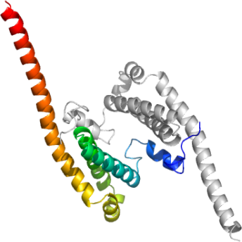
USP8 Dimerization Domain
PDB:2A9U
Revision
Revision Type:created
Revised by:created
Revision Date:created
Entry Clone Accession:gi:41281375
Entry Clone Source:MGC
SGC Clone Accession:usp08.0001.0142; plate SDC057:G11
Tag:N-terminal His-tag with integrated thrombin-cleavage site MGSSHHHHHHSSGLVPRGS.
Host:E.coliBL21 (DE3)
Construct
Prelude:
Sequence:gsMPAVASVPKELYLSSSLKDLNKKTEVKPEKISTKSYVHSALKIFKTAEECRLDRDEERAYVLYMKYVTVYNLIKKRPDFKQQQDYFHSILGPGNIKKAVEEAERLSESLKLRYEEAEVRKKLEEKDRQEEAQRLQQKRQETG
Vector:p28a-LIC
Growth
Medium:TB
Antibiotics:
Procedure:USP8 was expressed in E. coli BL21 (DE3) grown in Terrific Broth (TB) in the presence of 50 µg/ml of kanamycin at 37ºC to an OD600 of 7.5. Cells were then induced by isopropyl-1-thio-D-galactopyranoside (IPTG), final concentration 0.05 mM, and incubated overnight at 15ºC. The culture was centrifuged and the cell pellets were collected and stored at -80ºC.
Purification
Procedure
Immobilized-metal Affinity Chromatography: An aliquot (20 µl) of clarified supernatant is reserved for later analysis by SDS-PAGE. To the rest of it, 6 mL of 50% suspension of TALON metal-affinity resin (BD Bioscience) in the lysis buffer is added (3 mL suspension per each 50-mL tube with the supernatant). The tubes are incubated for 1 h in refrigerator with constant stirring, centrifuged at 4°C for 2 min at 1,000 rpm in SX4750 rotor using Allegra X-12R centrifuge (Beckman Coulter) and the supernatant is discarded. The affinity resin pellets are suspended in 10 mL lysis buffer and transferred into an Econo-Column (Bio-Rad 732-1010). The buffer is allowed to drain and discarded. The settled gel is washed with 15 mL lysis buffer containing 10 mM imidazol (washing buffer) followed by 15 mL washing buffer containing 0.05% Tween 20 (Sigma P7949) and 30 mL washing buffer; the washings are discarded. Elution is achieved using lysis buffer containing 200 mM imidazol. First 0.5 mL eluate is discarded and two 5-mL fractions are collected after that in tubes containing 50 µl 1M DTT (final DTT concentration is 10 mM). Protein concentration in these fractions is determined using Coomassie Plus Protein Assay reagent (Pierce 1856210) and bovine serum albumin (Pierce 23209) as a protein standard for calibration. The recombinant protein is usually eluted in the first 5-mL fraction and it is used directly at the next purification step. Some His-tagged proteins spread between the two fractions. In this case, the fractions are combined and concentrated down to approx. 5-mL volume by ultrafiltration (Amicon Ultra-15 10,000 MWCO, UFC901024 or 5,000 MWCO, UFC900524, as appropriate, Millipore).
Size-exclusion Chromatography: This step is performed using an AKTA Purifier or an AKTA Express system (GE Healthcare). The protein sample is loaded onto an XK16x65 column packed with HighLoad Superdex 200 (GE Healthcare) and equilibrated with 20 mM Tris-HCl, pH 8.0, 500 mM NaCl, 5% glycerol, 10 mM dithiothreitol (BioShop Canada, DTT 001). Elution is performed with the same buffer at a flow-rate of 3 mL/min in the Peak Fractionation mode: {Slope; min. peak width 0.833 min; level 0.000 mAU; peak start slope 10.000 AU/min; peak end slope 20.000 AU/min}. Note: If target protein has OD280 (0.1%, 1 cm) lower than 0.2, monitor the eluate absorbance at 215 ηm and use either time/volume-based or manual fraction collection. Fractions corresponding to major peak on the chromatogram are combined and analyzed by SDS-PAGE and LC/MS. Protein concentration is determined by measuring UV absorbance of the combined fractions at 280 ηm. Purified protein is concentrated using an ultrafiltration concentrator as above to a final concentration of approx. 50 mg/mL for further crystallographic screening (preceded by a PC test, Hampton Research, in order to determine optimal protein concentration) and biophysical characterization. The concentrated protein solution is divided into 100-µl aliquots that are frozen by immersion in liquid nitrogen and stored at -80ºC.
His-tag Removal: The purified protein, 10-20 mg, is diluted in 50 mM Tris-HCl, pH 7.5, 140 mM NaCl to a final volume of 4 mL and thrombin (Sigma T9681) is added (1 unit per mg target protein). The reaction mixture is incubated for 2 h at 21-23°C, and the protein is re-purified using size-exclusion chromatography as above.
Extraction
Procedure
The cell pellet was resuspended in lysis buffer (10 mM Tris-HCl, pH 8.0, 0.5 M NaCl, 5% glycerol, 2 mM imidazole, 1 mM ß-mercaptoethanol) inhibitor (0.1mM phenylmethyl sulfonyl fluoride, PMSF) and lysed using Microfluidizer. The lysate was cleared by centrifugation.
Concentration:
Ligand
MassSpec:
Crystallization:Purified USP8 was crystallized using the hanging drop vapor diffusion method. Crystals grew when the protein (12 mg/mL) was mixed with the reservoir solution in a 1:1 volume ratio, and the drop was equilibrated against a reservoir solution containing 27% PEG3350, 0.2 M lithium sulphate, 0.1 M bistris, 1 mM DTT at pH 5.6 in 293K temperature.
NMR Spectroscopy:
Data Collection:
Data Processing: