MAP3K3
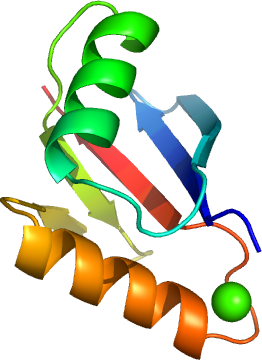
PDB:2C60
Revision
Revision Type:created
Revised by:created
Revision Date:created
Entry Clone Accession:gi|4505153
Entry Clone Source:MGC
SGC Clone Accession:Tag:mhhhhhhssgvdlgtenlyfq*s(m) TEV-cleavable (*) N-terminal his6 tag.
Host:BL21 (DE3)
Construct
Prelude:Sequence:mhhhhhhssgvdlgtenlyfq*sMGHSNRQSDVRIKFEHNGERRIIAFSRPVKYEDVEHKVTTVFGQPLDLHYMNNELSILLKNQDDLDKAIDILDRSSSMKSLRILLLSQD
Vector:pLIC- SGC1
Growth
Medium:Antibiotics:Procedure:Competent BL-21 (DE3) phage resistant cells were freshly transformed plasmid DNA and plated out onto LB plate plus 50 µg/mL kanamycin. A single colony was used to prepare a glycerol stock of the expression host and was frozen after over night growth using a glycerol concentration of 60 % in the cell suspension. The stock was stored at -80°C.
The glycerol stock was used to inoculate 10 mL TB media containing 50 µg/mL kanamycin in order to produce an overnight starting culture. The large scale growth was carried out at 37°C until approximately 30 min before induction when the temperature was lowered to 25°C. Protein production was induced with the addition of 1mM IPTG. Cells were grown for 12 hours at 25°C and were harvested by centrifugation at 4000 rpm for 15 minutes. The pellet was then stored in the -80°C freezer.
Purification
ProcedureColumn 1: Ni-affinity chromatography
Buffers: Affinity Binding Buffer: 10mM Imidazole, 300mM NaCl, 50mM pH8.0 NaH2PO4, 0.5mM TCEP. Affinity Wash Buffer: 50mM Imidazole, 300mM NaCl, 50mM pH8.0 NaH2PO4, 0.5mM TCEP. Affinity Elution Buffer: 250mM Imidazole, 300mM NaCl, 50mM pH8.0 NaH2PO4, 0.5mM TCEP.
Procedure: The cell extract was loaded on the column at 0.8 mL/min on an AKTA-express system (GE/Amersham). The column was then washed with 10 column volumes of Affinity Binding Buffer, 10 column volumes of Affinity Wash Buffer, and then eluted with Affinity Elution Buffer at 0.8 mL/min. The eluted peak of A280 was automatically collected.
Column 2: Gel filtration, Hiload 16/60, S75 16/60 - 120 mL
SEC-Buffers: 10 mM HEPES, pH 7.5, 500 mM NaCl, 5% glycerol, 0.5 mM TCEP.
Procedure: The eluted fractions from the Ni-affinity Histrap columns were loaded on the gel filtration column in GF buffer at 1.0 mL/min. Eluted proteins were collected in 1 mL fractions. 10 mM DDT was added to the purified protein before and after concentration. Using a Centricon 10 K cutoff concentrator the MAP3K3B-c012 pooled fractions was concentrated to 6.46 mg/mL. Concentration was determined from the absorbance at 280 nm.
Protein concentration: Centricon with a 10kDa cut off in SEC-buffer
Extraction
ProcedureExtraction buffer used: 10mM Imidazole, 300mM NaCl, 50mM pH8.0 NaH2PO4, 0.5mM TCEP, 1x complete PI EDTA free tablet/50mL.
The pellet (33.97 g) was resuspended with 3x volume of extraction buffer by placing the pellet in a 37°C water bath and vortexing. Once resuspended the cells were (1) broken by one passage through the Constant Systems cell breaker; (2) sonicating; (3) DNA precipitation with the addition of PEI to a final oncentration of 0.15 % for 30 mins on ice followed by a 17,000 rpm at 4°C to remove precipitation; (4) the supernatant was filtered through a GF/0.2 m M serum acrodiscs.
Concentration:
Ligand
MassSpec:
Crystallization:Crystals were obtained using the vapor diffusion method by mixing 100 nl of the concentrated protein (30 mg/mL) with 100nl of a well solution containing 0.2M NaI, 20% PEG3350 and 10 Ethylene glycole. Plate like crystals appeared after 3 days at 4°C.
NMR Spectroscopy:
Data Collection:
Data Processing: