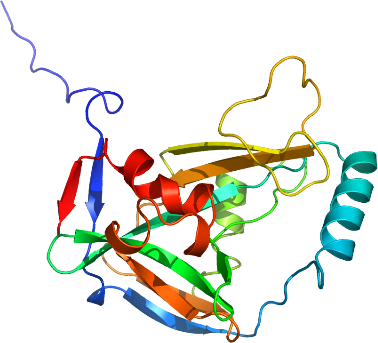
CPSF5
PDB:2CL3
Revision
Revision Type:created
Revised by:created
Revision Date:created
Entry Clone Accession:BC001403
Entry Clone Source:MGC
SGC Clone Accession:Tag:N-terminal hexahistidine tag with integrated TEV protease cleavage site: mhhhhhhssgvdlgtenlyfq*s(m).
Host:BL21(DE3)
Construct
Prelude:Sequence:MHHHHHHSSGVDLGTENLYFQSMGNKYIQQTKPLTLERTINLYPLTNYTFGTKEPLYEKDSSVAARFQRMREEFDKIGMRRTVEGVLIVHEHRLPHVLLLQLGTTFFKLPGGELNPGEDEVEGLKRLMTEILGRQDGVLQDWVIDDCIGNWWRPNFEPPQYPYIPAHITKPKEHKKLFLVQLQEKALFAVPKNYKLVAAPLFELYDNAPGYGPIISSLPQLLSRFNFIYN
Vector:pNIC-Bsa4
Growth
Medium:Antibiotics:Procedure:Cells from glycerol stocks were grown in 20 mL of TB supplemented with 8 g/L glycerol, 50 µg/mL Kanamycin at 30°C over night. The following morning 20 ml of the over night cultures inoculated 1500 mL Terrific Broth with trace elements, 4mM MgSO4, 18 g/L 87 % glycerol , 50 µg/mL kanamycin, 100 µL BREOX and in glass flasks in the Large Scale Expression System (LEX). Cells were grown at 37°C until OD600nm of 3. The cultivations were down-tempered to 18 °C for 1h in water bath. Expression of target protein was induced by addition of 0.5 mM IPTG and was allowed to continue over night at 18 °C.
Purification
ProcedureColumns:HiTrap Chelating 1 ml (IMAC); HiLoad 16/60 Superdex 75 Prep Grade (Gel filtration)
Purification was conducted automatically on an ÄKTA xpress system operated by UNICORN software at a flow of 0.8 ml/min. Prior to purification columns were equilibrated with IMAC Bind/Wash1 Buffer (HisTrap HP) and Gel filtration buffer (Superdex 75). The protein sample was loaded on the HisTrap HP column that was washed with IMAC Bind/Wash1 Buffer followed by IMAC Wash2 Buffer. Bound protein was eluted from the IMAC columns with 7.5 ml of IMAC Elution Buffer and loaded onto the gel filtration column. The chromatogram from Gel filtration showed one major protein peak that mainly consisted of CPSF5A-h001 as shown by SDS-PAGE analysis. TCEP was added to the pooled protein peak to a final concentration of 2 mM. The protein was concentrated to 13 mg/ml and stored at -80ºC.
Extraction
ProcedureCells were harvested by centrifugation and pellets were in 50mM Sodium-Fosfate pH 7.5, 500mM NaCl, 10% glycerol (IMAC Bind/Wash1 Buffer) supplemented with two tablets Complete EDTA-free protease inhibitor tablet and freezed at -80 ºC.The cells were briefly thawed in warm water and 2000 U of Benzonase was added. Disruption of cells was performed by sonication for 3 minutes (pulsed 1s-2s, on-off) and samples were centrifuged for 20 minutes at 40 000×g. The soluble fraction was decanted and filtered through 0.45µm prior to loading onto the ÄKTAxpress for further purification.
Concentration:LigandMassSpec:Crystallization:Sitting drops containing 0.1 µl of protein solution (13 mg/ml) + 0.1µl well solution containing 0.2M Calcium chloride, 0.1M MES pH 6, 20% w/v PEG 6000 was left to equilibrate against well solution. Crystals grew over several weeks. Seeding into pre-equilibrated drops was necessary to obtain crystals of the selenomethionine labeled protein.
NMR Spectroscopy:Data Collection:Data Processing:Native data to 1.9 Å was collected at BESSY, beamline BL14.1. The space group is P3121. We were able to produce small crystals of selenomethionine labelled protein by seeding. These diffracted to 2.5 Å, a SAD dataset was collected at ESRF ID14.2. This data had nice statistics to 3 Å, this resolution was used when running SOLVE. Three out of five selenium sites in the asymmetric unit was found. ARP/wARP was used to build the initial model using the high resolution native data. Repeated rounds of manual building using Coot and refinement using Refmac produced the final model which starts at glycine 21 and ends at the C-terminus of CPSF5, which is asparagine 227. Residues 134-138 was disordered and are not a part of the final model.