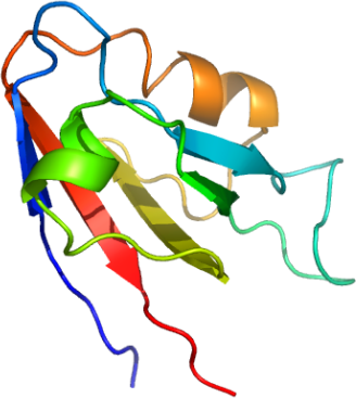
DLG3-PDZ domain
PDB:2FE5
Revision
Revision Type:created
Revised by:created
Revision Date:created
Entry Clone Accession:DLG3A-s001
Entry Clone Source:Origene
SGC Clone Accession:Tag:N-terminal hexahistidine tag
Host:BL-21(DE3)R3 phage resistant strain
Construct
Prelude:Sequence:mhhhhhhssgvdlgtenlyfq*smTIMEVNLLKGPKGLGFSIAGGIGNQHIPGDNSIYITKIIEGGAAQKDGRLQIGDRLLAVNNTNLQDVRHEEAVASLKNTSDMVYLKVAKPGS
Vector:pNIC28-Bsa4
Growth
Medium:Antibiotics:Procedure:Freshly transformed E. coli cells was used to inoculate 40 ml of LB containing 50 µg/ml kanamycin for overnight growth. The following day, 10 mls of this starter was used to inoculate 1 litre of TB plus 50 µg/ml kanamycin. When OD600 reached ~1.6 the temperature was shifted down from 37°C to 25°C for 1 hour before induction with the addition of 1 mM IPTG. Protein expression was allowed to carry on for a futher 4 hours before harvest. The cells were harvested by centrifugation, resuspended in lysis buffer before storing in a -80°C freezer.
Lysis/Binding Buffer: 50 mM Hepes pH 8.0, 500 mM NaCl, 5 % glycerol, 5 mM imidazole pH 8.0, 0.5 mM TCEP.
Purification
ProcedureColumn 1 : Wash Buffer: 50 mM Tris pH 8.0, 500 mM NaCl, 5 % Glycerol, 25 mM Imidazole pH 8.0, 0.5 mM TCEP; Elute Buffer: 50 mM Hepes pH 8.0, 500 mM NaCl, 5 % Glycerol, 250 mM Imidazole pH 8.0, 0.5 mM TCEP; GF Buffer: 50 mM Tris pH 8.5, 500 mM NaCl, 0.5 mM TCEP.
Procedure: The supernatent was filtered through a 1.2 micron and then a 0.2 micron syringe filter. 4 mls of a 50 % Ni2+-NTA slurry was added to a clean 15 mm diameter gravity column. The resin was equilibrated with 5 column volumes (10 ml) Lysis/Binding buffer. The supernatant was allowed to drip through the resin twice. The resin was then washed with 5 column volumes of Lysis/Binding Buffer and 25 column volumes of Wash Buffer. Finally the protein was eluted with 6 column volumes of Elute buffer and the eluate collected in 2ml fractions into eppendorf tubes.
Enzymatic treatment : HIS- TAG removal. To improve the purity of the sample the hexahistidine tag was cleaved and the cleavage products plus the contaminants rebound to a Ni2+-resin thus allowing the cleaved DLG3A sample to run through the column.
The DLG3A-p032 fractions were pooled and cleaved overnight with 100 µl of 2.8 mg/ml of in-house produced TEV protease. 500 µl of 50 % Ni-sepharose (Amersham) was added and the sample inverted repeatedly on a rotator for 60 minutes at 4°C to remove the His tag with TEV and other contaminants. The protein was concentrated to a final concentration of 143 mg/ml before storing at -80°C.
Extraction
ProcedureTo the cell pellet (approx. 80 mls) was added 20 mls of Lysis/Binding buffer and PMSF was added to a final concentration of 1.0 mM. Cell breakage: 4 passes through the Emulsiflex C5 high pressure homogeniser. Total vol: 100 mls (estimate).PEI was added to a final concentration of 0.05 % to precipitate the DNA . Centrifuge for 40 mins at 18000 rpm and 4°C to remove cell debris. Discard pellet.
Concentration:LigandMassSpec:Crystallization:Column 1 : Wash Buffer: 50 mM Tris pH 8.0, 500 mM NaCl, 5 % Glycerol, 25 mM Imidazole pH 8.0, 0.5 mM TCEP; Elute Buffer: 50 mM Hepes pH 8.0, 500 mM NaCl, 5 % Glycerol, 250 mM Imidazole pH 8.0, 0.5 mM TCEP; GF Buffer: 50 mM Tris pH 8.5, 500 mM NaCl, 0.5 mM TCEP.
Procedure: The supernatent was filtered through a 1.2 micron and then a 0.2 micron syringe filter. 4 mls of a 50 % Ni2+-NTA slurry was added to a clean 15 mm diameter gravity column. The resin was equilibrated with 5 column volumes (10 ml) Lysis/Binding buffer. The supernatant was allowed to drip through the resin twice. The resin was then washed with 5 column volumes of Lysis/Binding Buffer and 25 column volumes of Wash Buffer. Finally the protein was eluted with 6 column volumes of Elute buffer and the eluate collected in 2ml fractions into eppendorf tubes.
Enzymatic treatment : HIS- TAG removal. To improve the purity of the sample the hexahistidine tag was cleaved and the cleavage products plus the contaminants rebound to a Ni2+-resin thus allowing the cleaved DLG3A sample to run through the column.
The DLG3A-p032 fractions were pooled and cleaved overnight with 100 µl of 2.8 mg/ml of in-house produced TEV protease. 500 µl of 50 % Ni-sepharose (Amersham) was added and the sample inverted repeatedly on a rotator for 60 minutes at 4°C to remove the His tag with TEV and other contaminants. The protein was concentrated to a final concentration of 143 mg/ml before storing at -80°C.
NMR Spectroscopy:
Data Collection:
Data Processing: