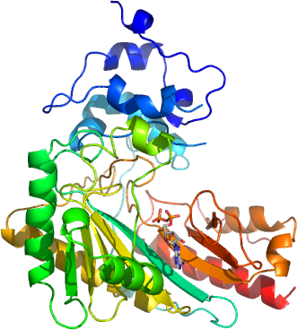
CKMT2 from sarcomeric
PDB:2GL6
Revision
Revision Type:created
Revised by:created
Revision Date:created
Entry Clone Accession:
Entry Clone Source:
SGC Clone Accession:
Tag:N-terminal histag with thrombin cleavage site: mgsshhhhhhssglvprgs
Host:E.coli BL21 (DE3)
Construct
Prelude:
Sequence:gsTTGYLLNRQKVCAEVREQPRLFPPSADYPDLRKHNNCMAECLTPAIYAKLRNKVTPNGYTLDQCIQTGVDNPGHPFIKTVGMVAGDEESYEVFADLFDPVIKLRHNGYDPRVMKHTTDLDASKITQGQFDEHYVLSSRVRTGRSIRGLSLPPACTRAERREVENVAITALEGLKGDLAGRYYKLSEMTEQDQQRLIDDHFLFDKPVSPLLTCAGMARDWPDARGIWHNYDKTFLIWINEEDHTRVISMEKGGNMKRVFERFCRGLKEVERLIQERGWEFMWNERLGYILTCPSNLGTGLRAGVHVRIPKLSKDPRFSKILENLRLQKRGTGGVDTAAVADVYDISNIDRIGRSEVELVQIVIDGVNYLVDCEKKLERGQDIKVPPPLPQF
Vector:p28a-LIC
Growth
Medium:
Antibiotics:
Procedure:10 µL competent BL-21 (DE3) codon + RIL cells were transformed with 1 µL plasmid for 20 min on ice followed by heatshock at 42degC for 1 min. 125 µL of SOC media was added to the cellsuspension which was incubated for 1 hour at 37degC and plated on LB-plates containing kanamycin (50 µg/mL) and chloromphenicol (25 µg/mL). 120 mL of LB medium with 50 µg/ml of kanamycin and 25 µg/ml of chloromphenicol was inoculated with colonies from the LB plate and incubated overnight (ON) at 37degC. After overnight, all of the seeds were inoculated into 1.8 L of Terrific Broth medium in the presence of 50 µg/ml of kanamycin and 25 µg/ml of chloromphenicol at 37ºC and grown to an OD600 of 4.0. Cells were then induced by isopropyl-1-thio-D-galactopyranoside at the final concentration of 1.0 mM and grown overnight at 18ºC in a LEX bubbling system.
Purification
Procedure
The supernatant was passed through DE52 (Whatman) column equilibrated with the binding buffer and then loaded onto 3 mL Ni-NTA column (Qiagen) equilibrated with the same binding buffer at 4 ºC. The Ni-NTA column was washed with 150 mL of the wash buffer (10mM Tris pH 7.5, 0.5 M NaCl, 5% glycerol, 30 mM imidazole) and the protein was eluted with 15 mL of the elution buffer (10mM Tris pH 7.5, 0.5 M NaCl, 5% glycerol, 250 mM imidazole). The protein was loaded on Superdex200 column (26x60) (Amersham Biosciences), equilibrated with 20 mM Tris-HCl pH 7.5, 0.5 M NaCl, 5 mM MgCl2, 5% glycerol, and 20mM DTT at flow rate 2.5 mL/min. 10 mM ADP, 10 mM creatine and 10 mM MgCl2 were added to the purified protein before concentration. The protein was concentrated using an Amicon Ultra centrifugal filter to the final concentration of 10 mg/mL. The protein concentration was measured using Bradford assay. About 7 mg of protein was obtained from 1.8 L of cell culture.
Extraction
Procedure
Cultures were centrifuged and the cell pellets were suspended in 100 ml of the binding buffer (10 mM Tris pH 7.5, 0.5 M NaCl, 5% glycerol, 5 mM imidazole) with a protease inhibitor cocktail (0.1 mM M benzamidine-HCl and 0.1 mM phenylmethyl sulfonyl fluoride) and flash frozen. The thawed cell pellet was lysed by a combination of 0.5% CHAPS (Sigma) and sonication. The lysate was centrifuged at 15000 rpm for 60 min and the supernatant was used for subsequent steps of purification.
Concentration:10 mg/mL
Ligand
MassSpec:
Crystallization:10 mM ADP, 10 mM creatine and 10 mM MgCl2 were added to the purified creatine kinase upon setting the crystallization trials using the sitting drop vapor diffusion method. The protein drop was equilibrated against a reservoir solution (1:1 volume ratio) containing 17-19 % PEG 3350, 0.2 M di-ammonium citrate, 10 mM DTT, and 100 mM Bis-Tris pH 6.0. Crystals reached a size of about 100 microns within five days after initially observing precipitation in the crystallization drops.
NMR Spectroscopy:
Data Collection:
Data Processing: