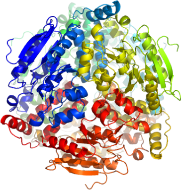
ECHS1
PDB:2HW5
Revision
Revision Type:created
Revised by:created
Revision Date:created
Entry Clone Accession:BC008906
Entry Clone Source:MGC
SGC Clone Accession:
Tag:TEV-cleavable (*) N-terminal his6 tag.
Host:BL-21(DE3)R3 phage resistant
Construct
Prelude:
Sequence:mhhhhhhssgvdlgtenlyfq*sMASGANFEYIIAEKRGKNNTVGLIQLNRPKALNALCDGLIDELNQALKIFEEDPAVGAIVLTGGDKAFAAGADIKEMQNLSFQDCYSSKFLKHWDHLTQVKKPVIAAVNGYAFGGGCELAMMCDIIYAGEKAQFAQPEILIGTIPGAGGTQRLTRAVGKSLAMEMVLTGDRISAQDAKQAGLVSKICPVETLVEEAIQCAEKIASNSKIVVAMAKESVNAAFEMTLTEGSKLEKKLFYSTFATDDRKEGMTAFVEKRKANFKDQ
Vector:pNIC28-Bsa4
Growth
Medium:
Antibiotics:
Procedure:Transformed 50 microL competent BL-21 (DE3) phage resistant cells with 10 µl of the plasmid DNA and plated out onto LB plate plus 50 mg/ml kanamycin. The next day colonies were picked out into fresh deep well blocks containing 1 ml TB + 50 mg/ml kanamycin. These were grown overnight and glycerol stocks prepared by adding 333 ml of 60 % glycerol to 1 ml of cell suspension, mixing and then storing in a -80°C freezer.
The glycerol stock was used to innoculate 10 mls of TB + 50 mg/ml kanamycin which was grown overnight at 37°C as a starter culture for a 1 litre growth. The large scale growth was grown at 37°C until approximately 30 mins before induction when the temperature was lowered to 18°C. Protein production was induced with the addition of 1mM IPTG. The next day cells were harvested by centrifugation at 4000 rpm for 15 minutes. The pellet was then stored in the -80°C freezer.
Purification
Procedure
Column 1 : Ni-affinity, HisTrap, 1 ml (GE/Amersham)
The cell extract was loaded on the column at 0.8 ml/min on an AKTA-express system (GE/Amersham). The column was then washed with 10 column volumes of Affinity Binding Buffer, 10 column volumes of Affinity Wash Buffer, and then eluted with Affinity Elution Buffer at 0.8 ml/min. The eluted peak of A280 was automatically collected.
Column 2 : Gel filtration, Hisload S75 16/60 - 120 ml
The eluted fractions from the Ni-affinity Histrap columns were loaded on the gel filtration column in GF buffer at 1.0 ml/min. Eluted proteins were collected in 1 ml fractions. 10 mM DTT were added to the purified protein before and after concentration.
Protein concentration: Using a Centricon 10 K cutoff concentrator the ECHS1A-c004 pooled fractions was concentrated to 13.8 mg/ml. Concentration was determined from the absorbance at 280 nm.
Extraction
Procedure
The pellet (22.33 gms) was resuspended with 3x volume of lysis buffer (approximately 100 mls final) by intermitently placing the pellet in a 37°C water bath and vortexing. Once resuspended the cells were (1) broken by one passage through the Constant Systems cell breaker; (2) sonicating; (3) DNA precipitation with the addition of PEI to a final oncentration of 0.15% for 30 mins on ice followed by a 17,000 rpm at 4 °C to remove precipitation; (4) the supernatant was filtered through a GF/0.2 serum acrodiscs.
Concentration:
Ligand
MassSpec:The expected mass with histidine is 31039. The experimentally determined mass was 31039.
Crystallization:Crystals grew from a 2:1 ratio mix of protein-to-reservoir: 0.2 M Na(form), 0.1M Bis-Tris propane,pH 8.5, 20 % PEG 3350, 10 % EtGly + 10mM MgCl2. Crystals were flash frozen directly in liquid nitrogen prior to data collection.
NMR Spectroscopy:
Data Collection:Resolution: 2.55 Å. X-ray source: Synchrotron SLS- SLS-X10 at a single wavelength 0.9791 Å.
Data Processing: