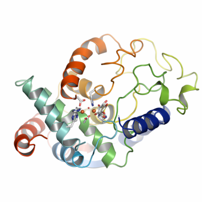
ALDRL6
PDB:2IBN
Revision
Revision Type:created
Revised by:created
Revision Date:created
Entry Clone Accession:BC073848
Entry Clone Source:SGC Clone Accession:Tag:Host:Bl21 (DE3)
Construct
Prelude:Sequence:SMDRVFTTYKLMHTHQTVDFVRSKHAQFGGFSYKKMTVMEAVDLLDGLVDESDPDVDFPNSFHAFQTAEGIRKAHPDKDWFHLVGLLHDLGKVLALFGEPQWAVVGDTFPVGCRPQASVVFCDSTFQDNPDLQDPRYSTELGMYQPHCGLDRVLMSWGHDEYMYQVMKFNKFSLPPEAFYMIRFHSFYPWHTGRDYQQLCSQQDLAMLPWVREFNKFDLYTKCPDLPDVDKLRPYYQGLIDKYCPGILSW
Vector:pNIC-Bsa4
Growth
Medium:Antibiotics:Procedure:30 µL competent BL21 (DE3) cells were transformed with 1 µL plasmid. Held 30min on ice, heat-shocked in 42 degree waterbath for 45sec at 42°C. Held on ice for 2 min. Added 100µL SOC and incubated in shaker for 1 h. Cells were plated on LA plates with 50 mg/l Kanamycin and 0.2% glucose. Glycerol stocks were made by adding 5 colonies to 1ml phosphate buffered TB with 50 mg/l Kanamycin. 150 ul culture was taken out at mid-logphase (3 hours growth at 37 degrees) and mixed with sterile glycerol to a final concentration of 20% and then frozen at -80.Glycerol stocks are used to inoculate 100 ml of LB + Kan (50 µg/ml). The cultures are shaken at 30 °C overnight. The overnight culture was used to inoculate 12 x 750 ml minimal media + Kan (50 µg/ml) (inoculation diluted 1/100). 5 hours later, OD 600 = 0.62, temperature is set to 18°C and an amino acid mix (including selenomethionine) is added.Followin 1 hour incubation, target expression was induced by addition of 0.5 mM IPTG. Expression performed at 18°C overnight.
Purification
ProcedureColumns:
- 1 mL Hi-Trap Chelating (Ni-charged). (GE Healthcare).
- Superdex 75 HiLoad 16/60 (GE Healthcare).
- 1 ml MonoQ (GE Healthcare).
The sample was purified using a peristaltic pump or ÄKTA-Purifier system (GE Healthcare). Briefly, sample was loaded on the IMAC column, eluted and loaded on the gel filtration column, eluted in a storage loop and then loaded on the gel filtration column. Elution fractions were pooled based on SDS-PAGE analysis. The N-terminal tag with the His-tag fusion and residual cloning elements were excised through incubation with tobacco etch virus protease (TEV; 1:20 protease:protein ratio) for 48 hours at 277K. After protease digestion, the IMAC purification was repeated to remove non-cleaved protein and the His-tagged TEV-protease. After additional purification by ion-exchange chromatography on a 1 ml MonoQ column (in Tris pH 8.0, 10 % glycerol by varying NaCl concentration), MIOX was essentially pure as judged by SDS-PAGE analysis.
Extraction
ProcedureCells were harvested by centrifugation (WCW 42 g) and pellets were resuspended in 80 ml of lysis buffer (50mM HEPES pH 7.5, 500mM NaCl, 10% glycerol, 10 mM Imidazole, 0.5 mm TCEP and 1 tablet Complete EDTA-free protease inhibitor (Roche Biosciences)). After adding 8 µl of a 250 U/µl benzonase (Novagen) stock solution the sample was sonicated (Sonics VibraCell) at 80% amplitude for 3 min (effective time, pulse: 4Â on 4Â off). The sample was spun for 30 min at 20500 rpm in a Sorvall SA-800 rotor and the soluble fraction decanted and filtered through a 0.45 µm flask filter.
Concentration:LigandMassSpec:Crystallization:MIOX crystals were grown by hanging drop vapor diffusion at 293° K. MIOX (15 mg/ml) in 20 mM Hepes pH 7.5, 300 mM NaCl, 10 % glycerol, 2 mM TCEP, 5 mM myo-inositol, 1 mM L-cysteine and 1 mM FeCl3 were mixed with an equal amount (0.6 µl) of reservoir solution (100 mM Tris pH 7.8, 1.8 M Ammonium sulphate). Crystals grew initially as rods (poorly ordered) but after two months incubation, plates appeared. The plates diffracted to 1.5 Å resolution at a synchrotron.
NMR Spectroscopy:Data Collection:A plate-shaped crystal of MIOX was used to collect single wavelength anomalous dispersion dataset was collected on the K-edge of selenium at the beam line ID29 (ESRF, Grenoble, France). Space group was C2 (119.7, 55.86, 111,5, ?=116.7°). XDS/XSCALE was used to process the data and XPREP/SHELXD/SHARP/PIRATE was used to solve the structure.
Data Processing: