Regulators of G-protein signalling (RGS) are domains that terminate the signal emanating from G-protein coupled receptors (GPCR). When a GPCR binds a specific ligand, a signal is generated that is transmitted to the G-proteins consisting of the Ga and Gbg subunits. In the inactive state Ga and Gbg exist as a complex with the Ga GTPase in an inactive GDP bound state. Upon activation, GDP is replaced with GTP which induces Ga and Gbg complex disassociation. At this stage both Ga and Gbg proteins are active. Ga does have an intrinsic GTPase activity but this is greatly enhanced by the association with an RGS domain. Hence RGS proteins act as GTPase activating proteins (GAPs). Once GTP hydrolysis has taken place the GDP-Ga protein can recombine with Gbg, thus terminating the signal.
RGS10 is a member of the D/R12 subfamily of RGS proteins. Unlike the R4 subfamily, RGS10 does not have an amphipathic helix at the N-terminus which for the R4 family members enhances the localisation of the protein to the membrane surface (Bernstein et al, 2000; Chen et al, 1999) where it is ideally placed to interact with the G-proteins. Instead, RGS10 is localised to the membrane surface by palmitylation of a conserved cysteine residue located in the RGS domain (Tu et al, 1999; Castro-Fernandez et al, 2002).
Phosphorylation of RGS10 by protein kinase C alpha causes its translocation to the nucleus with the result that its Galpha GAP activity is regulated through spatial reorganisation (Rimler et al, 2006).
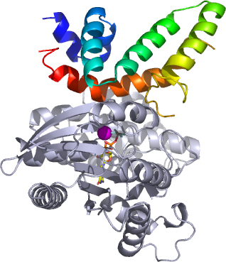
Here we present the crystal structure of RGS10 in complex with the activated state of Galphai3