BACH1
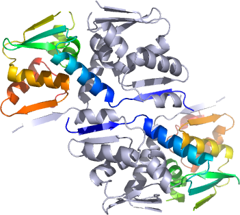
PDB:2IHC
Revision
Revision Type:created
Revised by:created
Revision Date:created
Entry Clone Accession:gi|45827690
Entry Clone Source:MGC
SGC Clone Accession:Tag: mhhhhhhssgvdlgtenlyfq*s(m) TEV-cleavable (*) N-terminal his6 tag.
Host:BL21 (DE3)
Construct
Prelude:Sequence:mhhhhhhssgvdlgtenlyfqsmSVFAYE SSVHSTNVLLSLNDQRKKDVLCDVTIFVE GQRFRAHRSVLAACSSYFHSRIVGQADGE LNITLPEEVTVKGFEPLIQFAYTAKLILS KENVDEVCKCVEFLSVHNIEESCFQFLKF
Vector:pNIC28-Bsa4
Growth
Medium:Antibiotics:Procedure:Starter cultures ( 10 ml LB, 50 µg/ml kanamycin) were inoculated with a single colony and grown overnight. LB media (1L) was inoculated with 5 ml of culture and grown at 37°C, 170 rpm until OD600 was 0.4. The flask was then transferred to an 18°C incubator and protein expression induced with 0.1 mM IPTG (final concentration) at an OD600 of approximately 0.5. Cells were harvested the following morning by centrifugation (10min, 6500 rpm, 4°C). The pellet was resuspended in 25 ml binding buffer and frozen at -20°C.
Purification
ProcedureColumns 1 & 2 : DE52 anion exchange resin/ IMAC Sepharose 6 Fast Flow resin, placed in series.
Gravity feed chromatography: DE52 anion exchange resin was prepared by suspending 10 g in 100 ml of 2.5 M NaCl. The resulting gel slurry was packed in a column and allowed to settle prior to equilibrating in approximately 50 ml of Binding Buffer. Nickel charged IMAC sepharose beads were prepared by packing 4 mls of gel slurry (approximately 50% beads in 30% EtOH) in a small column and equilibrating in approximately 10 ml of Binding Buffer. Both columns were then placed in series and the protein containing supernatant passed through under gravity. This was followed by washing using 50 ml of Binding Buffer followed by 50 ml of Wash Buffer. Elution of the protein proceeded using a stepwise gradient of elution buffer: 5 mls x 30 mM imidazole, 5 ml x 50 mM imidazole and 10 ml x 250 mM imidazole.
Enzymatic treatment: Protein containing fractions were pooled and the His-tag cleaved using TEV protease (reaction proceeded overnight at 4°C).
Column 3: Gel Filtration, HiLoad 16/60 Superdex 75 prep grade column run on an AKTA-prime system.
The protein solution was concentrated using a Vivaspin 20 ultrafiltration device with a 5 kDa MWCO to about 10 ml. A pre-equilibrated gel filtration column was injected with 5 ml of the solution and two runs undertaken at a flow rate of 0.8 ml/min.
Concentration: Using Vivaspin 5 ultrafiltration spin columns with a 5 kDa MWCO the protein was dialysed against 50 mM HEPES, pH 7.5, 100 mM NaCl, 0.5 mM TCEP and concentrated to 5 mg/ml approximated using UV absorbance at 280 nm and a predicted extinction coefficient of 4720.
Extraction
ProcedureThe cell pellet was defrosted and AEBSF plus CHAPS were added to a final concentration of 1 mM and 0.5% (w/v) respectively. Cells were lysed by sonication, 10 s on, 2 s off at 40% amplitude for 8 min. The lysate was cleared by centrifugation for 45 minutes at 21,500 rpm, 4°C.
Concentration:LigandMassSpec:Following TEV treatment of the protein mass spec confirmed the correct mass of 14,042 Da expected for this construct.
Crystallization:Crystals were obtained using the vapor diffusion method and a protein concentration of 10 mg/ml by mixing 750nl of the concentrated protein with 750nl of a well solution containing 20% PEG3350, 0.2M MgCl2, 0.1M Tris pH 8.5. Crystals appeared after several days at 20°C.
NMR Spectroscopy:Data Collection:Crystals were cryo-protected using the well solution supplemented with an additional 15% ethylene glycol and flash frozen in liquid nitrogen. Diffraction data were collected at the SLS beam line X10 ( l =0.979 Å) to 2.45Å
Data Processing: