PIM2: Human Proviral Integration Site for MuLV
PDB:2IWI
Revision
Revision Type:created
Revised by:created
Revision Date:created
Entry Clone Accession:GI:42821111
Entry Clone Source:Ben Turk (pEBG-Pim2)
SGC Clone Accession:Tag:mhhhhhhssgvdlgtenlyfq*sm (* TEV protease cleavage)
Host:E.coli BL21(DE3)R3
Construct
Prelude:Sequence:mhhhhhhssgvdlgtenlyfqsmLTKPLQ GPPAPPGTPTPPPGGKDREAFEAEYRLGP LLGKGGFGTVFAGHRLTDRLQVAIKVIPR NRVLGWSPLSDSVTCPLEVALLWKVGAGG GHPGVIRLLDWFETQEGFMLVLERPLPAQ DLFDYITEKGPLGEGPSRCFFGQVVAAIQ HCHSRGVVHRDIKDENILIDLRRGCAKLI DFGSGALLHDEPYTDFDGTRVYSPPEWIS RHQYHALPATVWSLGILLYDMVCGDIPFE RDQEILEAELHFPAHVSPDCCALIRRCLA PKPSSRPSLEEILLDPWMQTPAEDVPLNP SKGGPAPLAWSLLP
Vector:pLIC
Growth
Medium:Antibiotics:Procedure:10 ml from a 50 ml overnight culture in LB, 100µg/ml ampicillin was used to inoculate 1 litre of LB medium containing 100µg/ml ampicillin. Cultures were grown at 37°C until they reached an OD600 of 0.4 and then cooled to 18°C. Expression was induced for 4 hours using 0.15 mM IPTG. The cells were harvested by centrifugation, transferred to 50 ml tubes, resuspended in 30 ml binding buffer, and frozen.
Purification
ProcedureColumn 1 : Ion exchange - Nucleic acid removal . DEAE cellulose (DE52, Whatmann), 10 g of resin in 2.5 x 20 cm column. The resin was hydrated in 2.5M NaCl, then washed with 50 ml binding buffer prior to loading the sample.
Buffers: Binding buffer
Procedure: Supernatant was applied at gravity flow, followed by a wash with 50 ml binding buffer. The column flow-through was collected.
Column 2 : Ni-affinity Ni-sepharose, 5 ml of 50% slurry in 1.5 x 10 cm column, equilibrated with binding buffer.
Buffers : Binding buffer: 50 mM HEPES pH 7.5, 500 mM NaCl, 5% Glycerol; Wash buffer: 50 mM HEPES pH 7.5, 500 mM NaCl, 20 mM Imidazole, 5% glycerol; Elution buffer: 50 mM HEPES pH 7.5, 500 mM NaCl, 50-250 mM Imidazole, 5% Glycerol.
Procedure: The flowthrough from column 1 was loaded by gravity flow on the Ni-sepharose column. The column was then washed with 50 ml wash buffer at gravity flow. The protein was eluted by gravity flow by applying 5-ml portions of elution buffer with increasing concentration of imidazole (50 mM, 100 mM, 150 and 250 mM); fractions were collected until essentially all protein was eluted. After elution DTT was added to a final concentration of 10 mM.
Enzymatic treatment : (Dephosphorylation and Histag cleavage) Samples containing PIM-2 were pooled and 200µg GST-lambda phosphatase and 100µg TEV protease added for overnight incubation at 4°C: protein solution contained elution buffer plus 10 mM DTT and 0.05 mM MnCl2.
Column 3: MonoQ ion exchange
Buffers: Buffer A: 50 mM HEPES pH 7.5; Buffer B: 50 mM HEPES pH 7.5, 1 M NaCl.
Procedure: Dephosphorylated PIM-2 was applied to a 1 ml MonoQ column in buffer A and eluted from the column by a linear gradient with buffer B. Homogeneous non-phosphorylated PIM-2 eluted at ~100 mM NaCl.
Column 4: Size Exclusion Chromatography
Buffers: 50 mM HEPES pH 7.5, 250 mM NaCl
Procedure: The monoQ elution was concentrated to ~2 ml and injected onto a HiLoad 16/60 S75 Gel Filtration column and ran at 1 ml/min. 1.8 ml fractions were collected.
Concentration : Samples containing unphosphorylated protein were pooled and concentrated in Centricons (10 kDa cut off) to 11 mg/ml. Dephosphorylation was monitored using LC- ESI MS-Tof.
Extraction
ProcedureThe frozen cells were thawed on ice and binding buffer (plus 1 mM PMSF, 0.5 mM TCEP, 50 mM L-Arg, 50 mM L-Glu) added to a final volume of 50 ml. Cells were lysed using a high pressure cell disruptor. The lysate was centrifuged at 16,500 RPM for 30 minutes and the supernatant collected for purification.
Concentration:LigandMassSpec:Masses of purified proteins were confirmed using an Agilent LC/MSD TOF system with reversed-phase HPLC coupled to electrospray ionisation and an orthogonal time-of-flight mass analyser. Proteins were desalted prior to mass spectrometry by rapid elution from a C3 column with a gradient of 5-95% acetonitrile in water with 0.1% formic acid. The purified PIM-2 protein was homogeneous and had an experimental mass of 34277 Da as expected from its primary structure.
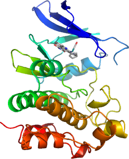
Crystallization:The ruthenium half-sandwich complex compound (R)- 5 was added to 0.6 mM (from a 10 mM stock solution in DMSO) prior to protein concentration to 11 mg/ml in 50 mM HEPES pH 7.5, 250 mM NaCl, 10 mM DTT. Crystals were grown at 4°C in 1.5µl sitting drops mixing 0.3µl PIM-2 solution with 1.2µl of mother liquor (90 mM HEPES pH 7.5, 1.44 M Na/KPO 4 ).
NMR Spectroscopy:Data Collection:PIM-2 complex with (R)- 5 (PDB 2IWI): 2.8 Å , X-ray source: Swiss Light Source synchrotron beamline SLS -X10.
Data Processing: