B56γ (PPP2R5C)
PDB:2JAK
Revision
Revision Type:created
Revised by:created
Revision Date:created
Entry Clone Accession:BC016183
Entry Clone Source:Mammalian Gene Collection
SGC Clone Accession:PPP2R5CA-k005
Tag:N-terminal hexahistidine tag with integrated TEV protease cleavage site: mhhhhhhssgvdlgtenlyfq*sm
Host:E.coli BL21(DE3) (Novagen)
Construct
Prelude:Sequence:mhhhhhhssgvdlgtenlyfq*smVVDAANSNGPFQPVVLLHIRDVPPADQEKLFIQKLRQCCVLFDFVSDPLSDLKWKEVKRAALSEMVEYITHNRNVITEPIYPEVVHMFAVNMFRTLPPSSNPTGAEFDPEEDEPTLEAAWPHLQLVYEFFLRFLESPDFQPNIAKKYIDQKFVLQLLELFDSEDPRERDFLKTTLHRIYGKFLGLRAYIRKQINNIFYRFIYETEHHNGIAELLEILGSIINGFALPLKEEHKIFLLKVLLPLHKVKSLSVYHPQLAYCVVQFLEKDSTLTEPVVMALLKYWPKTHSPKEVMFLNELEEILDVIEPSEFVKIMEPLFRQLAKCVSSPHFQVAERALYYWNNEYIMSLISDNAAKILPIMFPSLYRNSKT
Vector:pNIC-Bsa4
Growth
Medium:Antibiotics:Procedure:Native proteinCells from a glycerol stock were grown in 50 ml LB supplemented with 50 µg/ml kanamycin at 37 ºC overnight. 10 ml of the overnight culture was used to inoculate 0.75 l LB supplemented with 8 g/l glycerol and 50 µg/ml kanamycin. The culture was grown in a TunAir flask at 37 ºC until OD600 reached ~1. The flask was down-tempered to 18 ºC over a period of 1 hour before expression of target protein was induced by addition of 0.5 mM IPTG. Expression was allowed to continue overnight and cells were harvested the following morning by centrifugation (5,500 x
g, 20 min, 4 ºC). The resulting cell pellet (22.4 g wet cell weight) was stored at -80 ºC.
SeMet labeled protein
Cells from glycerol stock were grown in 100 ml LB supplemented with 50 µg/ml kanamycin at 30 ºC overnight. 80 ml of the overnight culture was used to inoculate 4 bottles with 1.5 l minimal medium (without amino acids) supplemented with 50 µg/ml kanamycin and approximately 100 µl BREOX FMT 30 anti-foam solution (Cognis Performance Chemicals UK Ltd). The culture was grown in a LEX bioreactor system (Harbinger Biotechnology) at 37 ºC until OD600 reached ~0.5. Amino acids were added (Van Duyne, G. D., J. Mol. Biol. 229, 105-124 (1993)) and the bottles were down-tempered to 18 ºC. Expression of target protein was induced one hour later by addition of 0.5 mM IPTG. The expression was allowed to continue at 18 ºC overnight. Cells were harvested the following morning by centrifugation (5,500 x g, 20 min, 4 ºC) and stored at -80 ºC.
Purification
ProcedureColumnsIMAC: Ni-charged 1 ml HiTrap Chelating HP (GE Healthcare)
Gel filtration column: HiLoad 16/60 Superdex 200 Prep Grade (GE Healthcare)
Procedure
Purification of the native protein was performed as a two step process on an ÄKTAxpress system (GE Healthcare). Prior to purification, columns were equilibrated with IMAC wash1 buffer and gel filtration buffer, respectively. The filtered lysate was loaded onto the Ni-charged HiTrap Chelating column and washed with IMAC wash1 buffer followed by IMAC wash2 buffer. Bound protein was eluted from the IMAC column with IMAC elution buffer and automatically loaded onto the gel filtration column. Fractions containing the target protein were pooled and fresh TCEP was added to a final concentration of 2 mM. The protein was subsequently concentrated using an Amicon Ultra-15 centrifugal filter device with 30,000 NMWL (Millipore) to 8.9 mg/ml in a volume of 3 ml and stored at -80 ºC. Purification of the SeMet enriched protein was performed in basically the same way. The filtered lysate was purified by IMAC and gel filtration chromatography followed by a concentration step. The final SeMet-protein concentration was 7.4 mg/ml. The identities of the proteins were confirmed by mass spectrometry and selenomethionine was fully incorporated at all 10 methionine positions in the labeled protein.
Extraction
ProcedureThe cell pellet was quickly thawed and resuspended in lysis buffer (1.5 ml/g cell pellet) supplemented with one tablet of Complete EDTA-free protease inhibitor (Roche Applied Science), 1000 U Benzonase (Merck) and a knife edge of lysozyme (Sigma). Cells were disrupted by high-pressure homogenization (TC5-0612W-332 from Stansted fluid power LTD) and cell debris was removed by centrifugation (49,000 x
g, 30 min, 4 ºC). The supernatant was decanted and filtered through a 0.45 µm flask filter. The SeMet-culture was treated in basically the same way.
Concentration:8.9 mg/mL
LigandMassSpec:The mass spectrometry analysis confirmed a mass of 45807 Da for the native protein and of 46270 for the selenomethionine labeled protein. All 10 methionine positions were fully incorportated by selenomethionine
Crystallization:Native crystals were obtained by the hanging drop vapour diffusion method using a 24-well plate. 2 µl of the protein solution (8.9 mg/ml) was mixed with 1 µl of well solution consisting of 0.1 M Tris pH 8.0, 20 mM L-proline and 29% MPD. The plate was incubated at 4 ºC and crystals were flash frozen in liquid nitrogen directly from the drop. SeMet crystals were produced in the same way by mixing 0.2 µl of the SeMet-protein solution (7.4 mg/ml) with 0.1 µl of well solution (the same as used for the native crystals).
NMR Spectroscopy:Data Collection:The native x-ray data was collected at station ID14.4 at ESRF. The selenomethionine data was collected at BM30 at ESRF.
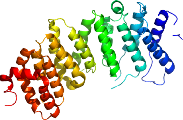
Data Processing:All data was processed using the programs XDS and XSCALE. The structure of B56y was solved by MAD phasing using Solve that was able to identify 7 of the 10 selenium sites present in the asymmetric unit. DM was used to perform phase extension of the 3.5 Å selenomethionine data to the 2.6 Å of the native data. The initial model was built in manually using coot. Refinement using refmac and further manual model building using coot gave the final model consisting of residues 30-372 of B56γ. Simulated annealing was done on the close to finished model to get rid of any potential model bias. The structure was refined to an R and Rfree of 21.6% and 26.0%, respectively. 99.7% of the residues of the model are within the most favored or additionally allowed regions of the ramachandran plot. The coordinates and structure factors were deposited in the PDB with accession code 2JAK.