JARID1C
PDB:2JRZ
Revision
Revision Type:created
Revised by:created
Revision Date:created
Entry Clone Accession:gi|11321605
Entry Clone Source:MGC
SGC Clone Accession:JARID1CA-c556
Tag:N-terminal, TEV cleavable (*) hexahistidine tag.
mhhhhhhssgvdlgtenlyfq(*)sm
Host:BL21(DE3)-R3-pRARE2
Construct
Prelude:Sequence:smNELEAQTRVKLNYLDQIAKFWEIQGSSLKIPNVERRILDLYSLSKIVVEEGGYEAICKDRRWARVAQRLNYPPGKNIGSLLRSHYERIVYPYEMYQSGANLVCNTRPFDNEEKDK
Vector:pNIC28-Bsa4.
Growth
Medium:Antibiotics:Procedure:BL21(DE3)-Rosetta competent cells were transformed with the expression plasmid and plated on LB plates containing 25 µg/ml kanamycin and 34µg/ml chloramphenicol.
5 Colonies from the transformation were used to inoculate 5 times 5 ml LB containing 25 µg/ml kanamycin and 34 µg/ml chloramphenicol. The cells were then grown at 250 RPM, 37°C up to OD 0.65 then induced with 1 mM IPTG for 3 h. Expression of the 5 clones was analyzed by SDS-PAGE. The best clone was used for large scale expression in either [
13C,
15N]-M9 medium, containing 0.5g [
15N]-NH4Cl and 2g [
13C]-glucose per L.
Starter overnight cultures of 2 x 50 ml were grown at 37°C in LB medium supplemented with 25 µg/ml kanamycin and 34 µg/ml chloramphenicol. The cells of 40 ml preculture were collected and resuspended in 10 ml M9 medium for inoculation of the large scale culture of 5 x 400 ml M9 with appropriate antibiotics in 5 x 2 L bottles (dilution for inoculation 1:80). The culture was grown at 37°C and transferred to 18°C when the OD600 reached a value of 0.6. The culture was induced with 0.5 mM IPTG and grown at 18°C overnight. The next day the cells were harvested by centrifugation, washed with ice-cold 150 mM NaCl and frozen at -80°C.
Purification
ProcedureColumn 1: Ni-affinity, MC-POROS, 8 ml (Applied Biosystems)
Column 2: Superdex 75, 320 ml
The cell extract was loaded on the column at 1 ml/min on a workstation Vision (Applied Biosystems). The column was washed with 10 column volumes of start buffer and eluted with a gradient from 5 to 500 mM imidazole at a flow rate of 5 ml/min. The extinction at 280 nm was monitored and fractions were collected and analyzed by SDS-PAGE. Positive fractions were pooled and filled into a dialysis bag with 3.5 kDa MW cutoff.
Dialysis and TEV-cleavage: The His-tag was cleaved with 1 mg TEV per 40 mg target protein in a dialysis bag and dialysed with 5 L of: 20 mM Tris HCl 8.0, 500 mM NaCl, 1 mM beta-mercaptoethanol, at 15°C overnight. The digest was checked by SDS-Page, as the cleavage was completed, the sample was supplemented with 5 mM arginine and glutamine concentrated to 3 ml on a 5 kDa Amicon Ultra-15 centrifuge concentrator. Fractions containing the protein of interest was collected and concentrated.
Extraction
ProcedureFrozen cell pellets from 2 L culture (ca. 6.3 g wet mass) were resuspended in a total volume of 25 ml lysis buffer. The cells were broken by 4 passes at 16,000 psi through a high pressure homogeniser (EmulsiFlex-C3/Avestin) followed by centrifugation for 60 minutes at 60,000g. The supernatant was further clarified by filtration (0.45 µm).
Concentration:Using Amicon Ultra-15 concentrators with 5 kDa cutoff, the JARID1CA-c556 was exchanged into NMR buffer and concentrated as follows: U-[
13C,
15N]- labelled JARID1CA: protein concentration 1 mM in 50 mM Phosphate, pH 6.0, 50 mM NaCl, 5 mM d DTT, 0,02% azide 10% D2O.
LigandMassSpec:Crystallization:NMR Spectroscopy:NMR spectra were acquired at 297 K, using Bruker DRX 600 and DMX 750 spectrometers in standard configuration with triple resonance probes equipped with self-shielded triple axis gradient coils. Spectra for the resonance and NOE assignment were recorded essentially as described in the original references. A 1 mM [
13C, [
15N-labelled sample of JARID1CA in 90% H2O/10% D2O (NMR buffer; pH 6.0) was used for all H N -detected triple resonance experiments, 3D CBCA(CO)NNH, CBCANNH, CC(CO)NNH, H(CCCO)NNH, HBHA(CBCACO)NNH, HNCO, HN(CA)CO, 3D
15N-separated NOESY-HSQC, [
15N T1 and
15N T2 relaxation, and heteronuclear
15N-
1H NOE experiments and for a 3D
13C-separated, aliphatic-centred NOESY-HSQC spectrum. The sample was then freeze-dried and re-dissolved in 100% D2O for acquisition of 3D
13C-separated HMQC-NOESY, HCCH-COSY, HCCH-TOCSY spectra. Data were processed using the program XWIN-NMR (version 2.6) of Bruker BioSpin GmbH ( Rheinstetten , Germany ).
Data Collection:Data Processing:Assignment of
13C,
15N and
1H resonances was carried out using standard assignment procedures on Silicon Graphics O2 workstations and an Intel Dual Xeon 3GHz PC, with the interactive program Sparky version 3.10 (
http://www.cgl.ucsf.edu/home/sparky/ ). The assignments are deposited in the BioMagResBank (
http://www.bmrb.wisc.edu/) under accession code BMRB- 15348
Assigned
1H-
15N-HSQC spectrum of the Bright/ARID domain from human JARID1CA recorded at 297 K and 750 MHz.
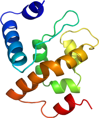
Preliminary three dimensional structures of the JARID1CA Bright/ARID domain were calculated using the program CYANA v. 2.0 ( Güntert et al., (1997) J. Mol. Biol. 273 , 283-298; Herrmann et al., (2002). J. Mol. Biol. 319 , 209-227 ) and were based on the resonance assignments together with NOE peak lists from
13C HMQC-NOESY, 3D
13C-aliphatic-centred NOESY-HSQC and 3D
15N NOESY-HSQC spectra. Dihedral angle restraints were predicted based on the chemical shift data using the program TALOS (Cornilescu et al., J. Biomol. NMR, 13 (1999) 289-302;
http://spin.niddk.nih.gov/NMRPipe/talos/) and were used to aid initial rounds of structure calculations. Most of these were excluded in the final rounds of calculations. A precise ensemble of structures showing the expected Bright domain fold was obtained. Hydrogen bond restraints were added on the basis of the protected amides following sample exchange into D2O. Iterative structure refinement was carried out using XPLOR-NIH v. 2.14 ( Schwieters et al., (2003) J. Magn. Reson. 160 , 66-74;
http://nmr.cit.nih.gov/xplor-nih/ ). Structures were refined in explicit water using CNS ( Linge et al (2003) Proteins, 50:496-506). Structures validation was performed using WHATIF
http://swift.cmbi.kun.nl/WIWWWI and the Protein Structure Validation Suite (PSVS)
http://www-nmr.cabm.rutgers.edu/PSVS . The refined ensemble of the 20 lowest energy structures with no NOE violations was submitted to the PDB under code 2JRZ. Chemical shift list and the list of NMR restraints were deposited with the structures.