NFATC2IP
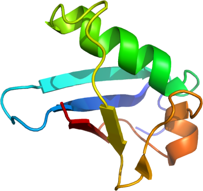
PDB: 2JXX
Entry Clone Accession: NP_116204.3
Entry Clone Source: ubh72.BC021551.OBS.40K16.pOTB7
SGC Clone Accession: ubh72.342.419 (SDC088D093)
Tag: N-terminal: MGSSHHHHHHSSGLVPRGS
Host: BL21 (DE3)
Vector:pET28-MHL
Sequence:MGSSHHHHHHSSGLVPRGSTETSQQLQLRVQGKEKHQTLEVSLSRDSPLKTLMSHYEEAMGLSGRKLSFFFDGTKLSGRELPADLGMESGDLIEVWG
Growth
Medium: M9 minimal media: 100 uM ZnSO4, 8.55 mM NaCl, 47.6 mM Na2HPO4, 22 mM KH2PO4, 100 mM MgSO4, 2 mM biotin, 1.5 mM thiamine.HCl, 10 mM ZnSO4, and 0.1 M CaCl2.
Procedure:A 125 ml flask containing M9 minimal media, supplemented with 15NH4Cl, 13C6-D-glucose, and 50 µg/ml kanamycin was inoculated from a glycerol stock of bacteria. The flask was shaken overnight (18 hours) at 220 rpm at 37 degC.A 2L flask containing 1000 ml minimal media supplemented with 50 µg/ml kanamycin was inoculated with the 50 ml overnight starter culture, and incubated at 37 degC to an OD600 of 1.0. Protein expression was induced with 100 µM isopropyl-thio-ß-D-galactopyranoside and the cells were incubated for overnight (15.5 hours) at 220rpm at 15 degC, and cell pellets collected by centrifugation and frozen in 50 mL Falcon tubes at -80 degC.
Purification
Procedure:
IMAC: The lysate was clarified by centrifugation for 20 min at 4 degC. The supernatant was mixed with 2 mL of Ni2+ affinity beads per 40 mL lysate. The mixture was incubated with mixing for 20 minutes at 4 degC. The lysate was spun at 2000 rpm for 6 minutes, and the supernatant was decanted. The remaining resin was resuspended and washed twice with lysis buffer, followed by two washes with with 5 mL of cold wash buffer. The washed resin was transferred to a gravity filter column and further washed with 2 mL of wash buffer. Samples were eluted from the resin by exposure to 5mL of elution buffer.
Buffer exchange & protein concentration: The purified protein was exchange from elution buffer into MOPS-based NMR buffer by ultracentrifugation using 2 mL concentrators with a 3,000 molecular weight cut-off (VivaSpin 2 MES) at 3000 rpm, resulting in a final volume of 300ul (final protein concentration of 0.9 mM). The concentrated protein was transferred to a 3mm NMR tube.
Residual dipolar coupling sample alignment: After data collection was performed on the unaligned sample, the purified protein was aligned by titrating 12 mg/ml Pf1 co-solvent Protease-free Phage into the NMR sample until 10 Hz proton splitting was observed.
Extraction
Procedure: The frozen cell pellet stored in a 50 ml Falcon tube obtained from 1L of culture was thawed by soaking in warm water. Each cell pellet was resuspended in 40 mL lysis buffer and lysed by sonication on ice.
Structure Determination
NMR Spectroscopy:A series of spectra (3D 1H-13C NOESY, 3D 1H-15N NOESY, 2D 1H-13C Constant Time HSQC, 3D HNCO, 3D HNCA, 3D CBCA(CO)NH, 3D HBHA(CO)NH, 3D 1H-13C Aromatic NOESY, 3D (H)CCH-TOCSY, and 3D H(C)CH-TOCSY) were generated using a 500MHz Bruker AVANCE spectrometer, a 600MHz Bruker AVANCE spectrometer and a 800MHz Bruker AVANCE spectrometer. Spectra of aligned and unaligned spectra (2D 1H-15N IPAP HSQC) were obtained using the 500MHz Bruker AVANCE spectrometer and the 800MHz Bruker AVANCE spectrometer. NMR data was processed and analyzed using Topspin, NMRPipe, NMRDraw, Sparky, Abacus, FMC-GUI, CNS, TALOS, PALES, PSVS, and WhatIF.