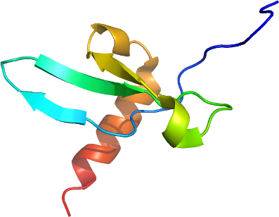
CBX4
PDB:2K28
Revision
Revision Type:created
Revised by:created
Revision Date:created
Entry Clone Accession:NP_003646
Entry Clone Source:Open Bios. LIFESEQ6297222 AmpR
SGC Clone Accession:
Tag:MGSSHHHHHHSSGLVPRGS
Host:BL21 (DE3)
Construct
Prelude:
Sequence:mgsshhhhhhssglvprgsEHVFAVESIEKKRIRKGRVEYLVKWRGWSPKYNTWEPEENILDPRLLIAFQNRERQEQ
Vector:p28a-thrombin-lic
Growth
Medium:
Antibiotics:
Procedure:A 250 ml flask containing M9 base minimum media (with 13C-glucose and 15N (NH4)2SO4) supplemented with 50 ug/ ml kanamycin (BioShop Canada KAN 201) was inoculated from a fresh transformed plate. The flask was shaken overnight (16 hours) at 250 rpm at 37 degC. Using the Lex system, a 2L bottle (VWR 89000-242) containing 1800 ml of minimum media supplemented 50 ug/ ml kanamycin and 600 ul antifoam 204 (Sigma A-8311) was inoculated with 50 ml overnight LB culture, and incubated at 37 degC. The temperature of the media was reduced to 15oC one hour prior to induction and induced at OD(600) = 2 with 100 uM isopropyl-thio-β-D-galactopyranoside (BioShop Canada IPT 001). Cultures were aerated overnight (16 hours) at 15 degC, and cell pellets collected by centrifugation and frozen at -80 degC.
Purification
Procedure
IMAC: Unclarified lysate was mixed with 2-3 mL of Ni-NTA superflow Resin (Qiagen) per 40 mL lysate. The mixture was incubated with mixing for at least 45 minutes at 4oC. The mixture was than loaded onto an empty comlum (BioRad) and washed with 100 ml wash buffer. Samples were eluted from the resin by exposure to 2-3 column volumes (approx. 10-15 mL) of elution buffer.
Gel filtration chromatography: An XK 26x65 column (GE Healthcare) packed with HighLoad Superdex 75 resin (GE Healthcare) was pre-equilibrated with gel filtration buffer for 1.5 column volumes using an AKTA explorer (GE Healthcare) at a flow rate of 2.5mL/min. The eluate sample from the IMAC step (approx. 15 mL) was loaded onto the column at 1.5 mL/min, and 2mL fractions were collected into 96-well plates (VWR 40002-012) using peak fractionation protocols). Fractions observed by a UV absorption chromatogram to contain the protein were pooled.
Protein concentration: Purified proteins were concentrated using 15 mL concentrators with a 5,000 molecular weight cut-off (Amicon Ultra-15, UFC900524, Millipore) at 3750 rpm, typically resulting in a final concentration of 4-5 mg/mL.
Extraction
Procedure
Frozen cell pellet contained in bags (Beckman 369256) obtained from 2L of culture were thawed by soaking in warm water. Each cell pellet was resuspended in 25-40 mL lysis buffer and homogenized using an Ultra-Turrax T8 homogenizer (IKA Works) at maximal setting for 30-60 seconds per pellet. Cell lysis was accomplished by sonication (Virtis408912, Virsonic) on ice: the sonication protocol was 10 sec pulse at half-maximal frequency (5.0), 10 second rest, for 10 minutes total sonication time per pellet.
Concentration:4-5 mg/mL.
Ligand
MassSpec:
Crystallization:
NMR Spectroscopy:All of the spectra were recorded at 25 °C on Varian INOVA 600 MHz and Bruker Avance 500 MHz spectrometers equipped with triple resonance 1H/13C/15N cold and cryoprobes, respectively. The final NMR samples contained 90% H2O, 10% D2O with a protein concentration ranging between 0.5 and 0.7 mM. The spectra were processed with NMRPipe software and analyzed with the SPARKY program (T. D. Goddard and D. G. Kneller, SPARKY 3, University of California, San Francisco). Linear prediction in the 13C and 15N dimensions was used to improve the digital resolution. Distance restraints for structure calculations were obtained from 13C-edited NOESY-HSQC (m = 120 ms) and 15N-edited NOESY-HSQC (m = 150 ms) experiments. NOESY spectra were analyzed with the SPARKY program. Structure calculations were carried out using CNS version 1.1 with its standard annealing protocol. The 20 lowest energy structures were refined using CNS by performing a short constrained molecular dynamics simulation in explicit solvent.
Data Collection:
Data Processing: