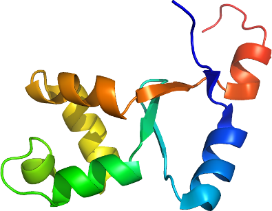
HERC2
PDB:2KEO
Entry Clone Accession:NP_004658
Entry Clone Source:herc2.BC070076.OBS.EHS10017571194.pBluescriptR
SGC Clone Accession:herc2.1203.1296.106D09 (SDC106D09)
Tag:N-terminal: MHHHHHHSSGRENLYFQ*G
Host:BL-21(DE3)
Vector:pET28MHL
Sequence: mhhhhhhssgrenlyfqgNNEKVTLVRIADLENHNNDGGFWTVIDGKVYDIKDFQTQSLTENSILAQFAGEDPVVALEAALQFEDTRESMHAFCVGQYLEPDQEGVTIPDLG
Growth
Procedure: A 250 ml flask containing M9 base minimum media (with 13C-glucose and 15N (NH4)2SO4) supplemented with 50 ug/ ml kanamycin (BioShop Canada KAN 201) was inoculated from a fresh transformed plate. The flask was shaken overnight (16 hours) at 250 rpm at 37 degC.
Using the Lex system, a 2L bottle (VWR 89000-242) containing 1800 ml of minimum media supplemented 50 ug/ ml kanamycin and 600 ul antifoam 204 (Sigma A-8311) was inoculated with 50 ml overnight LB culture, and incubated at 37degC. The temperature of the media was reduced to 15 degC one hour prior to induction and induced at OD600 = 2 with 100 uM isopropyl-thio-ß-D-galactopyranoside (BioShop Canada IPT 001). Cultures were aerated overnight (16 hours) at 15 degC, and cell pellets collected by centrifugation and frozen at -80 degC.
Purification
Procedure: Unclarified lysate was mixed with 2-3 mL of Ni-NTA superflow Resin (Qiagen) per 40 mL lysate. The mixture was incubated with mixing for at least 45 minutes at 4 degC. The mixture was than loaded onto an empty comlum (BioRad) and washed with 100 ml wash buffer. Samples were eluted from the resin by exposure to 2-3 column volumes (approx. 10-15 mL) of elution buffer.
Gel filtration chromatography: An XK 26x65 column (GE Healthcare) packed with HighLoad Superdex 75 resin (GE Healthcare) was pre-equilibrated with gel filtration buffer for 1.5 column volumes using an AKTA explorer (GE Healthcare) at a flow rate of 2.5mL/min. The eluate sample from the IMAC step (approx. 15 mL) was loaded onto the column at 1.5 mL/min, and 2mL fractions were collected into 96-well plates (VWR 40002-012) using peak fractionation protocols). Fractions observed by a UV absorption chromatogram to contain the protein were pooled.
Protein concentration. Purified proteins were concentrated using 15 mL concentrators with a 5,000 molecular weight cut-off (Amicon Ultra-15, UFC900524, Millipore) at 3750 rpm, typically resulting in a final concentration of 4-5 mg/mL.
Extraction
Procedure: Frozen cell pellet contained in bags (Beckman 369256) obtained from 2L of culture were thawed by soaking in warm water. Each cell pellet was resuspended in 25-40 mL lysis buffer and homogenized using an Ultra-Turrax T8 homogenizer (IKA Works) at maximal setting for 30-60 seconds per pellet. Cell lysis was accomplished by sonication (Virtis408912, Virsonic) on ice: the sonication protocol was 10 sec pulse at half-maximal frequency (5.0), 10 second rest, for 10 minutes total sonication time per pellet.
Structure Determination
NMR Spectroscopy:A series of spectra (3D 1H-13C NOESY, 3D 1H-15N NOESY, 3D 1H-13C_Aromatic NOESY, 2D 1H-13C Constant Time HSQC, 3D HNCO, 3D HNCA, 3D CBCA(CO)NH, 3D HBHA(CO)NH, 3D (H)CCH-TOCSY, and 3D H(C)CH-TOCSY) were generated using 800MHz Bruker AVANCE and 600MHz Varian INOVA spectrometers. NMR data was processed and analyzed using NMRPipe, NMRDraw, and Sparky programs. FMC-GUI was used for sequence specific resonance assignment. Structure was calculated and refined using CYANA, TALOS, and CNS.