OTUD7A
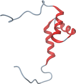
PDB:2L2D
Entry Clone Accession:OpenBiosystems cDNA template (IMAGE5729413)
Entry Clone Source:
SGC Clone Accession:otu04.0011-0083.136C12 (SDC136C12)
Host:Bacteria
Vector:pET28MHL vector (GenBank, EF456735)
Sequence: MHHHHHHSSGRENLYFQGSAECWAALLHDPMTLDMDAVLSDFVRSTGAEPGLARDLLEGKNWDLTAALSDYEQLRQVHTANLPHVFNEGRG
Growth
Medium:M9 minimal media supplemented with biotin, thiamine, and 10 µM ZnSO4; 15NH4Cl and 13C-glucose were the sole nitrogen and carbon source
Procedure:Bacteria were grown in M9 minimal media supplemented with biotin, thiamine, and 10 µM ZnSO4; 15NH4Cl and 13C-glucose were the sole nitrogen and carbon source. Starter cultures (50 mL in a 250 mL flasks) were prepared with media supplemented with 100 µL of glycerol stock and shaken overnight (18 hours) at 220 rpm at 37 ºC. They were used to inoculate 500 mL of growth media in a modified LEX fermentation system at 37 ºC to an OD600 of 1.0. Cultures were induced with 1 mM IPTG and grown at room temperature overnight (15.5 hours). Cells were harvested by centrifugation and frozen in 50 mL Falcon tubes at -80 ºC.
Purification
Procedure: Lysate was clarified by centrifugation for 20 min at 4C and the supernatant was mixed for 20 minutes at 4C with 2 mL settled Ni2+ affinity beads. Beads were batch-washed twice with 5 mL of cold wash buffer (spun at 2000 rpm for 6 minutes), transferred to a column, and further washed with 2 mL of wash buffer. Protein was eluted with 5 mL of elution buffer. Buffer exchange into NMR buffer (for H2O experiments): (10 mM Tris-HCl, 300 mM NaCl, 10 mM DTT, 1 mM Benzamidine, 0.01% NaN3, 1x inhibitor cocktail (Roche), 0.01 mM ZnSO4, 10% D2O, and 90% H2O; pH 7.0); (for D2O experiments): (10 mM Tris-HCl, 300 mM NaCl, 10 mM DTT, 1 mM Benzamidine, 0.01% NaN3, 1x inhibitor cocktail (Roche), 0.01 mM ZnSO4, and 100% D2O; pH 7.0) and protein concentration was performed using VivaSpin concentrators with a 5,000 molecular weight cut-off at 3000 rpm, resulting in a final volume of 300 ul. Protein samples were transferred to a 5 mm shigemi NMR tube for data collection.
Extraction
Procedure: Frozen cell pellets from 500 ml cultures were thawed, resuspended in 25 mL lysis buffer and lysed by sonication.
Structure Determination
NMR Spectroscopy:The NMR experiments were carried out at 25ºC on either Bruker Avance 600 or 800 MHz spectrometers equipped with cryogenic probes. All 3D spectra employed a non-uniform sampling scheme in the indirect dimensions and were reconstructed by multi-dimensional decomposition (MDD). The backbone assignments were obtained using HNCO, CBCA(CO)NH, HBHA(CO)NH, HNCA, and 15N-edited NOESY-HSQC spectra. Assignments were made with the automated method FAWN using FMCGUI program. Aliphatic side chain assignments relied on (H)CCH-TOCSY and H(C)CH-TOCOSY spectra. Aromatic ring resonances were assigned using 3D 13C-edited NOESY spectra. Stereospecific valine and leucine methyl assignments were obtained on the basis of the 13C-13C one-bond couplings in a high resolution 2D 1H-13C HSQC spectrum of 7%- 13C, 100%- 15N OTUD7A_11_83. Distance restraints for structure calculations were derived from cross-peaks in 15N-edited NOESY-HSQC (Tm = 100 ms) and 13C-edited aliphatic and aromatic NOESY-HSQC in H2O (Tm = 100 ms), respectively. NOE peaks were picked and integrated with the program SPARKY. Automated NOE assignment and structure calculations were performed using the program CYANA, version 2.1. A total of 86 phi and psi torsion angle restraints were derived from the program TALOS+. Hydrogen bond restraints were applied only for residues that were clearly in the secondary structure regions as judged by NOE patterns and chemical shifts and supported by TALOS+. The quality of the CYANA calculation was assessed by NMR structure quality assessment scores (NMR RPF scores) The best 20 of 100 CYANA structures from the final cycle were subjected to restrained molecular dynamics simulation in explicit water by the program CNS. The final structures were inspected by PROCHECK and MolProbity using the NESG validation software package PSVS.
Data Processing:The NMR structure of the UBA domain of OTUD7A has been deposited on 2010.08.17 into the RCSB database with PDB ID 2L2D and the following PDB authors: Wu B., Yee, A., Lemak, A., Gutmanas, A., Houliston S., Semesi A., Montelione, G., Dhe-Paganon., S. and Arrowsmith C.H.