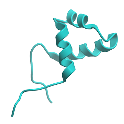
DMTF1
PDB:2LLK
Revision
Revision Type:created
Revised by:created
Revision Date:created
Entry Clone Accession:AT87-E10
Entry Clone Source:MGC
SGC Clone Accession:jmc21d06
Tag:MHHHHHHSSGRENLYFQG
Host:BL21 (DE3) cells (Invitrogen, C6000-03)
Construct
Prelude:
Sequence:MHHHHHHSSGRENLYFQGDRNHVGKYTPEEIEKLKELRIKHGNDWATIGAALGRSASSVKDRCRLMKDTCNTG
Vector:pET28-MHL
Growth
Medium:M9 minimal media (100 uM ZnSO4, 8.55 mM NaCl, 47.6 mM Na2HPO4, 22 mM KH2PO4, 100 mM MgSO4, 2 mM biotin, 1.5 mM thiamine.HCl, 10 mM ZnSO4, and 0.1 M CaCl2) supplemented with 15NH4Cl, 13C6-D-glucose, and 50 μg/ml kanamycin
Antibiotics:kanamycin
Procedure:Competent BL21 (DE3) cells (Invitrogen, C6000-03) were transformed and incubated overnight (18 hours) at 220 rpm at 37 °C in a 125 ml flask containing 50 ml of M9 minimal media (100 uM ZnSO4, 8.55 mM NaCl, 47.6 mM Na2HPO4, 22 mM KH2PO4, 100 mM MgSO4, 2 mM biotin, 1.5 mM thiamine.HCl, 10 mM ZnSO4, and 0.1 M CaCl2) supplemented with 15NH4Cl, 13C6-D-glucose, and 50 μg/ml kanamycin. The overnight starter culture was transferred to a 2 L flask containing 1 L of M9 minimal media supplemented with 15NH4Cl, 13C6-D-glucose, and 50 μg/ml kanamycin, and incubated at 37 °C. When the OD(600) reached a value of 1.0, protein expression was induced with 100 μM isopropyl-thio-β-D-galactopyranoside and the cells were incubated overnight (15.5 hours) at 220rpm at 15°C . Cell pellets were collected by centrifugation (7000 rpm, 20 mins) and frozen in 50 mL Falcon tubes at -80°C for storage
Purification
Procedure
The supernatant was mixed with 2 mL of Ni2+ affinity beads per 40 mL lysate. The mixture was incubated with mixing for 20 minutes at 4°C . The lysate was spun at 2000 rpm for 6 minutes, and the supernatant was decanted. The remaining resin was resuspended and washed twice with lysis buffer, followed by two washes with with 5 mL of cold wash buffer (15.4mM tris.HCl, 100 uM ZnSO4 100uL, 0.5 mM NaCl, and 30 mM imidazole; pH 8.5). The washed resin was transferred to a gravity filter column and further washed with 2 mL of wash buffer. Samples were eluted from the resin by exposure to 5mL of elution buffer (15.4mM tris.HCl, 100 uM ZnSO4 100uL, 0.5 mM NaCl, and 500 mM imidazole; pH 8.5).
Extraction
Procedure
The frozen cell pellet stored in a 50 ml Falcon tube obtained from 1L of culture was thawed by soaking in warm water, and resuspended in 40 mL lysis buffer (15.4mM tris.HCl, 100 uM ZnSO4 100uL, 0.5 mM NaCl, and 15 mM imidazole; pH 8.5). The cell pellet was lysed by sonication (Branson Sonicator) on ice for 10 minutes total sonication time (10 sec pulses at half-maximal frequency with 10 second rest)]. The lysate was clarified by centrifugation for 20 min at 4°C .
Concentration:
Ligand
MassSpec:
Crystallization:
NMR Spectroscopy:The purified protein was exchange from elution buffer into phosphate-based NMR buffer (for H2O experiments: 10 mM sodium phosphate, 300 mM NaCl, 1 mM Benzamidine, 0.01% NaN3, 0.01 mM ZnSO4, 10% D2O, and 90% H2O, pH 7.0; for 10 mM Tris-HCl, 400 mM NaCl, 1 mM Benzamidine, 0.01% NaN3, 0.01 mM ZnSO4, and 100% D2O, pH 7.0) by ultracentrifugation using 5 mL concentrators with a 5,000 molecular weight cut-off (VivaSpin 2 MES) at 3000 rpm, resulting in a final volume of 300ul (final protein concentration of 0.9 mM). The concentrated protein was transferred to a 5mm Shigemi NMR tube.
Data Collection:A series of spectra (3D 1H-13C NOESY, 3D 1H-15N NOESY, 2D 1H-13C Constant Time HSQC, 3D HNCO, 3D HNCA, 3D CBCA(CO)NH, 3D HBHA(CO)NH, 3D 1H-13C Aromatic NOESY, 3D (H)CCH-TOCSY, and 3D H(C)CH-TOCSY) were generated using a 600MHz Bruker AVANCE spectrometer and a 800MHz Bruker AVANCE spectrometer. NMR data was processed and analyzed using Topspin, NMRPipe, NMRDraw, MDD-GUI, Sparky, Abacus, FMC-GUI, CYANA, CNS, TALOS, PALES, PSVS.
Data Processing: