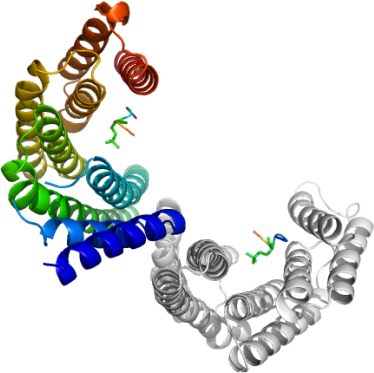
Cp 14-3-3 ε
PDB:2NPM
Revision
Revision Type:created
Revised by:created
Revision Date:created
Entry Clone Accession:cgd3_1290 (CryptoDB.org)
Entry Clone Source:Cp gDNA Iowa strain
SGC Clone Accession:Tag:N-Terminal hexahistidine tag with integrated tobacco etch virus (TEV) protease cleavage site.
Host:E. coli BL21 (DE3) Rosetta-R3
Construct
Prelude:Sequence:mgsshhhhhhssgrenlyfqgILRTQHTIRDSELNIKNSKMSDSVNARESNVYMAKLAEQAERYDEMAKYMKDVVEARQESEELTVEERNLLSVAYKNAVGSRRSSWRIISSVEQKEHSRNAEDASKMCGKYRSKVEAELTDICNDILTMLDKHLIPTATSPDSKVFYFKMKGDYHRYISEFSTGDSKQSSAEDALKAYKDATVVAKDLEPTHPIRLGLALNFSVFHYEILNEPRAAIDMAKEAFEMAIEQLDKLSEDCYKDSTLIMQLLRDNLTLWTAD
Vector:p15-TEV-LIC
Growth
Medium:Antibiotics:Procedure:Cryptosporidium parvum putative 14-3-3 protein was expressed in E. coli BL21 (DE3) Oxford in Terrific Broth (TB) in the presence of ampicillin and chloramphenicol (100 µg/mL and 34 µg/mL respectively). A single colony was inoculated into 50 mL of LB with ampicillin and chloramphenicol in a 125 mL flask and incubated with shaking at 250 rpm overnight at 37 ºC. The culture was transferred into 2 X 1.8 L TB with ampicillin and chloramphenicol and 0.3 mL of antifoam (Sigma) in 2 L bottles and cultured using the LEX system to an OD600 of 4.0. The culture was cooled to 15 ºC, and isopropyl-1-thio-D-galactopyranoside (IPTG) was added to 0.4 mM, and the culture was incubated overnight at 15 ºC.
Purification
ProcedureColumn 1: The cleared cell lysate was loaded onto a column containing 10 g DE-52 resin (Whatman), and then directly onto a 2.5 mL Ni-NTA (Qiagen) column at approximately 1.5 mL/min. When all the lysate was loaded, the two column system was washed with 15 mL binding buffer. The Ni-NTA column was then washed with 200 mL of Wash Buffer (50 mM HEPES pH 7.5, 500 mM NaCl, 30 mM imidazole, and 5 % glycerol) at 2 mL/min. After washing, the protein was eluted from the Ni-NTA column with 15 mL of Elution Buffer (50 mM HEPES pH 7.5, 500 mM NaCl, 250 mM imidazole, and 5 % glycerol). EDTA was added immediately to 1 mM; and DTT was added to 1 mM 15 minutes later.
TEV Protease Cleavage: The eluted Cp 14-3-3 protein was treated with TEV protease in a 1:500 molar ratio of protease:protein, and incubated at 4°C overnight concurrent with dialysis against 10 mM HEPES, pH 7.5, 500 mM NaCl, and 5 mM imidazole. The cut protein was separated from the uncut protein by passage through another 2.5 mL Ni-NTA column.
Column 2: The cut Cp 14-3-3 was applied to a Sephadex S200 26/60 gel filtration column (GE Healthcare) pre-equilibrated with 10 mM HEPES, pH 7.5 and 500 mM NaCl. The collected fractions corresponding to the eluted protein peak were concentrated to 10 mg/mL using a 15 mL Amicon Ultra centrifugal filter device (Millipore). Aliquots of the purified Cp 14-3-3 protein were stored at -70 °C.
Extraction
ProcedureThe culture was harvested by centrifugation and the cell pellet was suspended in 160 mL of binding buffer (50 mM HEPES, pH 7.5, 500 mM NaCl, 5% glycerol, and 5 mM imidazole) with protease inhibitor (1 mM benzamidine-HCl and 1 mM phenylmethyl sulfonyl fluoride, PMSF) and kept in 50 mL Falcon tubes at  80 ºC. Before purification, the cell suspension was thawed overnight at 4 ºC. Prior to mechanical lysis, each tube of cell suspension was pretreated with 0.5 % CHAPS and 500 units of benzonase (per 40 mL of resuspended cell pellet) for 40 minutes at room temperature. Then the cells were mechanically lysed with a microfluidizer (Microfluidizer Processor, M-110EH) at approximately 18000 psi. The lysate was centrifuged at 24000 rpm for 20 minutes at 10 ºC.
Concentration:10 mg/mL
LigandMassSpec:Crystallization:Purified Cp 14-3-3 was first treated with 2 mM DTT and a three fold molar excess of consensus peptide 2  RAI(pS)LP where pS denotes a phosphoserine  followed by incubation on ice for 1 h. The protein:peptide complex was crystallized using the hanging drop vapour diffusion method in a 24-well Linbro plate. The protein solution (1 µL) was added to 1 µL reservoir buffer containing 14% PEG3350, 0.1 M Calcium Acetate, 0.2 M trimethylamine-N-oxide, and 0.1 M HEPES pH 7.0, placed over 500 µL reservoir buffer, and incubated for at 293 K. Diamond shaped crystals (100-200 µm) grew to maximum size in two days.
NMR Spectroscopy:Data Collection:Data Processing: