ASS
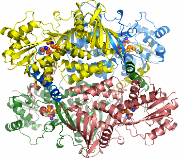
PDB:2NZ2
Revision
Revision Type:created
Revised by:created
Revision Date:created
Entry Clone Accession:Entry Clone Source:SGC Clone Accession:Tag:Host:Construct
Prelude:Sequence:SMSSKGSVVLAYSGGLDTSCILVWLKEQGYDVIAYLANIGQKEDFEEARKKALKLGAKKVFIEDVSREFVEEFIWPAIQSSALYEDRYLLGTSLARPCIARKQVEIAQREGAKYVSHGATGKGNDQVRFELSCYSLAPQIKVIAPWRMPEFYNRFKGRNDLMEYAKQHGIPIPVTPKNPWSMDENLMHISYEAGILENPKNQAPPGLYTKTQDPAKAPNTPDILEIEFKKGVPVKVTNVKDGTTHQTSLELFMYLNEVAGKHGVGRIDIVENRFIGMKSRGIYETPAGTILYHAHLDIEAFTMDREVRKIKQGLGLKFAELVYTGFWHSPECEFVRHCIAKSQERVEGKVQVSVLKGQVYILGRESPLSLYNEELVSMNVQGDYEPTDATGFININSLRLKEYHRLQSKVTAK
Vector:pNIC-Bsa4
Growth
Medium:Antibiotics:Procedure:30 microL competent BL-21 (DE3) cells were transformed with 2 microL plasmid miniprep for 30 min on ice followed by heatshock at 42°C for 45 sec. SOC, 125 µl, was added to the cellsuspension which was then incubated for 1 hour at 37°C and plated on LB-plates containing kanamycin (50 µg/mL). 30 mL TB with 100 µg kanamycin/ml was inoculated with cells and grown overnight (ON) at 30°C. Six x 7.5 ml of the inoculation culture was added to six TunAir flasks (Shelton Scientific) containing 750 mL of phosphate TB with 50 µg kanamycin/mL. Cultures were incubated at 37°C until OD600 reached 1. At this time the incubation temperature was lowered to 18°C and expression of ASS was induced by addition of 0.5 mM IPTG and continued for 18 hours. Cells were harvested by centrifugation in a SLC-6000 rotor for 10 minutes at 5000 rpm (WCW 70 g). Pellets were suspended in 140 mL 50 mM Naphosphate pH 7.5, 10 % glycerol, 0.5 mM TCEP and 300 mM NaCl, 10mM imidazole and Complete EDTA-free protease inhibitor (Roche Biosciences). Suspended cells were stored at -80°C until further use. Before lyses, 12 µl of 250U/µl benzonase (Novagen) was added to the suspended cells and the sample was sonicated (Sonics VibraCell) at 80% amplitude for 3 min (pulse: 4Â on and 4Â off).The sample was spun for 30 min at 20500 rpm in a Sorvall SA-800 rotor. The soluble fraction was decanted and filtered through a 0.45 µm syringe filter
Purification
ProcedureThe cleared lysate was loaded onto a His Trap Crude column by using a peristaltic pump and affinity-bound protein was eluted from the column on an Äkta prime by sequentiallyincreasing the imidazole concentration. The protein eluted at 260 mM imidazole and was further purified by gel filtration on a Superdex 200 gel filtration column in Gel filtration buffer. Fractions containing protein were pooled, the TCEP-concentration adjusted to 2 mM, and concentrated to 1.86 mg/ml in amicon filters
TEV-cleavage: TEV-cleavage was performed by adding a of 1.27 µm TEV-protease (final concentration) to the protein solution (38 µM) and letting it incubate for approximately 12 hours at 4degC. The sample was diluted with 30 mM Hepes, 500 mM NaCl, 10% glycerole and 0.5 mM TCEP, pH 7.5 before loading it onto a His-trap crude column on an Äkta Prime. The cleaved protein was detected in the flow-through. After elution the TCEP concentration was adjusted to 2 mM and the sample concentrated to 32 mg/ml in an AmiconUltra MWCO 10000 concentrator.
Extraction
ProcedureConcentration:LigandMassSpec:Crystallization:Crystals of Argininosuccinate synthase were found in several conditions during initial screening. However, well diffracting and useful crystals could only be obtained using seeding techniques. This was done by mixing equal amounts of protein solution at a concentration of 15 mg/ml containing 10mM Aspartate and 10mM Citrulline and reservoir solution containing 16% PEG 3350 and 0.15M DL-Malic Acid pH 7.0. After one day incubation the drops were streak-seeded from the initial crystal hits. Crystals appeared after 4-6 days and had reached their maximal size in ten days (approximately 300 micrometer × 300 micrometer × 50 micrometer).
As cryoprotectant, 25% Glycerol was added to the drop, after then crystals were harvested and flash-freezed in liquid nitrogen.
NMR Spectroscopy:
Data Collection:Diffraction data was collected at beamline 14.1 at the BESSY synchrotron radiation facility in Berlin, Germany. All data were indexed and integrated in space group F222 with the XDS package. The cell parameters are a=95.94 Å, b=117.47 Å, c=155.15 Å.
Data Processing:The structure was solved by Molecular Replacement using the Thermotoga maritima Argininosuccinate synthase structure as search model (PDB entry: 1VL2). The asymmetric unit contains one protein monomer. Refmac was used for refinement and Coot for model building. In the refinement, data in the interval 19.42-2.40Å resolution was used and the progress of refinement was followed by decreasing R and Rfree values. At the end of the refinement the values for R= 0.1975 and Rfree= 0.2647. The final model starts at lysine 4 and ends at serine 407. Residues 1-3, 156, 377 and 408-412 are disordered and not visible in the electron density map. A Ramachandran plot shows 89.3 % of all residues in favored regions and no outliers in the disallowed regions. Coordinates for the crystal structure were deposited in the Protein Data Bank, accession code 2NZ2.