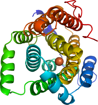
Plasmodium vivax Ribonucleotide reductase, small subunit R2 (Pv-RNR-R2)
PDB:2O1Z
Revision
Revision Type:created
Revised by:created
Revision Date:created
Entry Clone Accession:Pv086155
Entry Clone Source:P. vivax Salvador I gDNA (kindly donated by John Barnwell of CDC)
SGC Clone Accession:Tag:N-terminal His-tag with integrated TEV protease site: MGSSHHHHHHSSGRENLYFQG
Host:BL21-(DE3)-Rosetta-Oxford
Construct
Prelude:Sequence:ADVLNISKIPIFSKKEKKFSDLQKSKEANEKILSKETDRFTLYPILYPDVWDFYKKAEASFWTAEEIDLSSDLKDFEKLNDNEKHFIKHVLAFFAASDGIVLENLASKFLRQVKITEAKKFYAFQIAVENIHSETYSLLIDNYIKDEKERMNLFHAIENIPAVKNKALWAAKWINDTNSFAERIVANACVEGILFSGSFCAIFWFKKQNKLHGLTFSNELISRDEGLHTDFNCLIYSLLENKLPEEVVQNIVKEAVEVERSFICESLPCDLIGMNSRLMSQYIEFVADRLLECLGSPKIFHAKNPFNWMDL
Vector:p15Tev-Lic
Growth
Medium:Antibiotics:Procedure:PV-R2 was expressed in E. coli BL21-(DE3)-Rosetta-Oxford cells in Terrific Broth (TB) in the presence of kanamycin/chloramphenicol (50 µg/mL and 25 µg/mL respectively). A single colony was inoculated into 10 mL of LB with of kanamycin/chloramphenicol (50 µg/mL and 25 µg/mL respectively) in a 125 mL flask and incubated with shaking at 250 rpm overnight at 37 ºC. The culture was transferred into 100 mL of TB with 50 µg/mL kanamycin in a 250 mL shaking flask and incubated at 37 ºC for 3 hours. The culture was transferred into 2 X 1.8 L TB with 50 µg/mL kanamycin and 0.3 mL of antifoam (Sigma) in 2 L bottles and cultured using the LEX system to an OD600 of 2.5. The culture was cooled to 15 ºC, and isopropyl-1-thio-D-galactopyranoside (IPTG) was added to 0.4 mM, and the culture was incubated overnight at 15 ºC.
Purification
ProcedureThe cleared lysate was loaded onto a column prepacked with 10 g DE52 (Whatman) anion exchange resin (previously activated with 2.5 M NaCl and equilibrated with Binding Buffer); and subsequently onto a 1.0 - 2.5 mL Ni-NTA (Qiagen) column pre-equilibrated with Binding Buffer at approximately 1 - 1.5 mL/min. The volume of the Ni-NTA resin was pre-determined by the predicted protein yield from test expression analysis. After the lysate was loaded, the DE52 was further washed with 20 mL of Binding Buffer( added 100ul Protease inhibitor). Each Ni-NTA column was then washed with 200 mL of Wash Buffer (50 mM HEPES pH 7.5, 500 mM NaCl, 30 mM imidazole, and 5 % glycerol) at 20 mL/min. After washing, the protein was eluted with 15 mL of Elution Buffer (50 mM HEPES pH 7.5, 500 mM NaCl, 250 mM imidazole, and 5 % glycerol). EDTA was immediately added to the elution fraction to 1 mM; and DTT was added to 5 mM after 15 minutes. The Ni-NTA purified protein was loaded onto a 26/60 S200 Superdex gel filtration column; and the major peak corresponding to the PV-RNR was collected then added 10micromolar FeSO4 and after that concentrated the sample using a 15 mL Amicon Ultra centrifugal filter device (Millipore). The concentrated protein was stored at 4C
His Tag cleavage:
40microL of in house purified TEV was added to 40 mg of your protein. 1mM DTT was added. The concentration of protein was diluted to 4 mg/mL by adding binding buffer, then kept at 4 degC overnight.set up Bio-Rad Econo (1.5 x 15 cm x 27 mL) columns with 1.5 mL of suspended Ni-NTA Superflow resin. Equilibrate the columns with 50 mL of Binding Buffer and let drain completely by gravity.Determine the percentage of His-tag cleavage from the molecular weights of the cut and uncut protein samples on the SDS gel or you can run mass spect. Apply the protein samples onto the equilibrated Ni-NTA Superflow resin ( 2ml) and collect the flow-through. Then add more Binding Buffer to each column and check by by assaying 10 microL of each sample in 100 microL of Bradford reagent. Add 10 mL of Wash Buffer if protein does not come off the column in Binding Buffer.
Extraction
ProcedureThe culture was harvested by centrifugation and the cell pellet was suspended in 160 mL of binding buffer (50 mM HEPES, pH 7.5, 0.5 M NaCl, 5% glycerol, and 15 mM imidazole) with protease inhibitor (1 mM benzamidine-HCl and 1 mM phenylmethyl sulfonyl fluoride, PMSF) and kept in 50 mL Falcon tubes at  80 ºC. Before purification, the cell suspension was thawed overnight at 4 ºC. Prior to mechanical lysis, each tube of cell suspension was pretreated with 0.5 % CHAPS and 500 units of benzonase (per 40 mL of resuspended cell pellet) for 40 minutes at room temperature. Then the cells were mechanically lysed with a microfluidizer (Microfluidizer Processor, M-110EH) at approximately 18000 psi. The lysate was centrifuged at 24000 rpm for 20 minutes at 10 ºC.
Concentration:5mg/ml
LigandMassSpec:Crystallization:Purified protein, with tag removed, was crystallized using the hanging drop vapor diffusion method in a 24-well plate. Mother liquor (500 microL) containing 31.5% PEG2K MME and 0.15M K thiocyanatewas added to the buffer reservoir, and 1 microL protein solution was mixed with 1 microL mother liquor, then the plate was kept at 18 degC. The crystals appeared overnight.
NMR Spectroscopy:Data Collection:Data Processing: