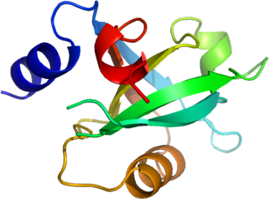
MPDZ : Human multiple PDZ domain protein (1st domain)
PDB:2O2T
Revision
Revision Type:created
Revised by:created
Revision Date:created
Entry Clone Accession:MPDZA_s001
Entry Clone Source:SGC Clone Accession:Tag:N-terminal hexahistidine tag before TEV cleavage site; C-terminal PDZ recognition motif.
Host:BL-21(DE3)R3 phage resistant
Construct
Prelude:sm highlights N-terminal residues present as a result of cloning procedure.
yykv C-terminal extension containing PDZ recognition motif
Sequence: smPACDEFDQLIKNMAQGRHVEVFELLKP PSGGLGFSVVGLRSENRGELGIFVQEIQE GSVAHRDGRLKETDQILAINGQALDQTIT HQQAISILQKAKDTVQLVIARGSLPQyyk v
Vector:pNIC28-Bsa4
Growth
Medium:Antibiotics:Procedure:Transformed 90 microL competent BL-21 (DE3) phage resistant cells containing the Rosetta plasmid with 2 µl of the plasmid DNA and plated out onto LB plate plus 50 µg/ml kanamycin and 34 µg/ml chloramphenicol. The next day colonies were picked out into 50 mls of LB + 50 µg/ml kanamycin + 34 µg/ml chloramphenicol and grown at 37°C overnight as a starter culture for a 1 litre growth. The large scale growth was grown at 37°C until approximately 30 mins before induction (OD600= 1.4) when the temperature was lowered to 25°C. Protein production was induced with the addition of 1mM IPTG. The next day cells were harvested by centrifugation at 4000 rpm for 15 minutes and resuspended in Lysis buffer before storage in the -80°C freezer. Lysis buffer: 10mM Imidazole, 300mM NaCl, 50mM pH8.0 NaH2PO4, 0.5mM TCEP, 1x complete PI EDTA free tablet/50mls.
Purification
ProcedureColumn 1: Low pressure chromatography using Bio-Rad Econo column (2.5 cm x 13 cm).
Procedure: 1ml of 50 % Ni-NTA slurry was added to a clean empty gravity column (providing a resin column of 0.5 ml volume). The resin was equilibrated with 10 mls of Lysis buffer. The supernatant was allowed to drip through the resin twice. The resin was then washed with 50 mls of Lysis Buffer follwed by 30 mls of Wash Buffer. Finally the protein was eluted with 15 mls of Elute buffer and the eluate collected in 2 ml fractions into eppendorf tubes.
Column 2: Gel filtration, Hiload 16/60, S75 16/60 - 120 ml
Procedure: The eluted fractions from the Ni-affinity Histrap columns were loaded on the gel filtration column in GF buffer at 1.0 ml/min. Eluted proteins were collected in 1 ml fractions.
Enzymatic treatment : At this stage the purity of the protein was greater than 95 % based on SDS-PAGE analysis. The C-terminal hexahistidine tag was removed by TEV protease treatment. The TEV protease, a hexahistidine-tagged construct, was over-expressed and purified in-house to a final concentration of 2.5 mg/ml. Add 30 µl of the TEV protease was added to each fraction and left at 4°C overnight. The following steps were carried out to remove the cleaved products and TEV protease. Place 200 µll of 50 % Ni-NTA agarose in a 1.5 ml eppendorf tubes, add 1ml of 50 mM Tris pH 8, 150 mM NaCl mix, spin down and remove buffer. Repeat this resin wash step once. Add the TEV treated protein sample to the resin and mix for 30 min. Finally spin down resin and collect the supernatant which contains the cleaved MPDZA@1. The sample was then concentrated to 3.5 mg/ml using a 10 kD cutoff spin concentrator before storage in a -80°C freezer.
Extraction
ProcedurePMSF 0.01M in 10 ml of Lysis/ Binding Buffer and added to the thawed cell pellet (approximately 40mls volume) and mixed by inversion. The cell pellet was lysed by passing it through the EmulsiFlex C5 high pressure homogeniser continuously for 10 minutes, collecting a final volume of approximately 50mls. PEI was added to a final concentration of 0.15% and the cell debris and precipitated DNA were spun down at 21,500rpm for 45 mins).
Concentration:LigandMassSpec:After His-tag removal - Expected MWt: 12886.6; Measured MWt: 12887.1.
Crystallization:Crystals grew from a 2:1 ratio mix of MPDZ 1st PDZ domain-to-reservoir (0.5% jeffamine 2001, 1.1M sodium malonate, pH 7.0) at 20°C.
NMR Spectroscopy:Data Collection:Resolution: Cryo: 20% ethylene glycol; 2.7 Å; X-ray source: synchrotron SLS -X10; single wavelength: 0.97945 Å
Data Processing:Method Used To Determine The Structure: molecular replacement; Software Used: PHASER; Starting Model: SWISSMODEL was used to generate a model based on sequence homology.