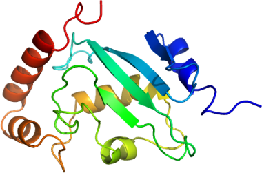
Pf UBC E2H: Plasmodium falciparum ubiquitin conjugating enzyme E2H
PDB:2ONU
Revision
Revision Type:created
Revised by:created
Revision Date:created
Entry Clone Accession:PF10_0330 (plasmodb.org)
Entry Clone Source:Synthetic DNA from Codon Devices
SGC Clone Accession:
Tag:N-Terminal hexahistidine tag with integrated tobacco etch virus (TEV) protease cleavage site.
Host:E. coli BL21 (DE3)-R3-pRARE2
Construct
Prelude:
Sequence:mhhhhhhssgrenlyfqgTSLTRKQCDFTKLIMAGYDLELNNGSTQDFDVMFHGPNGTAYEGGIWKVHVTLPDDYPFASPSIGFMNKLLHPNVDEASGSVCLDVINQTWTPLYSLVNVFEVFLPQLLTYPNPSDPLNSDAASLLMKDKNIYEEKVKEYVKLYASKDLWE
Vector:
Growth
Medium:TB
Antibiotics:100 microG/mL ampicillin and 34 microG/mL chloramphenicol
Procedure:A single colony was inoculated into 10 mL of LB with of Antibiotics and incubated with shaking at 250 rpm overnight at 37 degC. The culture was transferred into 50 mL of TB with Antibiotics in a 250 mL shaking flask and incubated at 37 degC for 3 hours. The culture was then transferred into 1.8 L of above-specified growth medium with Antibiotics and 0.3 mL of antifoam (Sigma) in a 2L bottle and cultured using the LEX system to an OD600 of ~5, cooled to 15 degC and induced with 0.5 mM isopropyl-1-thio-D-galactopyranoside (IPTG) overnight at 15 degC.
Purification
Procedure
The cleared cell lysate was loaded onto a column containing 10 g DE-52 resin (Whatman) anion exchangeresin (previously activated with 2.5 M NaCl and equilibrated with Binding Buffer), and then directly onto a 2.5 mL Ni-NTA (Qiagen) column at approximately 1.5 mL/min. When all the lysate was loaded, both columns were washed with 15 mL binding buffer. The Ni-NTA column was then washed with 200 mL of Wash Buffer at 2 mL/min. After washing, the protein was eluted from the Ni-NTA column with 15 mL of Elution Buffer. EDTA was added immediately to 1 mM.DTT was added to 5 mM 15 minutes later.
The eluted protein was applied to a Sephadex S200 26/60 gel filtration column (GE Healthcare) pre-equilibrated with gel filtration buffer.The fractions corresponding to the eluted protein peak were collected.
The purified protein was treated overnight at 4degC with TEV protease in a 1:200 molar ratio of protease:protein with the addition of 2 mM DTT and 15 mM imidazole. The cut protein was separated from the uncut protein by passage through another 2.5 mL Ni-NTA column.The buffer of the cut protein was exchanged to Crystal Buffer and the protein was concentrated to 15 mg/mL using an Amicon 5,000 MW cutoff spin concentrator.
Extraction
Procedure
Cells were resuspended to approximately 40 mL/L of cell culture in Binding Bufferwith protease inhibitor (1 mM benzamidine-HCl and 1 mM phenylmethyl sulfonyl fluoride, PMSF). Resuspended pellets stored at -80 degC were thawed overnight at 4 degC on the day before purification. Prior to mechanical lysis, each pellet from 1 L of culture was pretreated with 0.5% CHAPS and 500 units of benzonase for 40 minutes at room temperature. Cells were mechanically lysed with a microfluidizer (Microfluidizer Processor, M-110EH) at approximately 18000 psi; and the cell lysate was centrifuged using at ~75000 x g for 20 minutes at 10 degC.
Concentration:15 mg/mL
Ligand
MassSpec:
Crystallization:The protein was first treated with 2 mM DTT and was then crystallized using the sitting drop vapour diffusion method in a 96-well IntelliPlate (Hampton Research). The protein solution (0.3 microL) was added to 0.3 µL reservoir buffer containing 25% PEG3350, 0.1 M ammonium sulfate and 0.1 M Tris pH 8.5, with 100 µL reservoir buffer in the reservoir, and incubated at 18degC. Diamond shaped crystals grew to maximum size in 5 days.
NMR Spectroscopy:
Data Collection:
Data Processing: