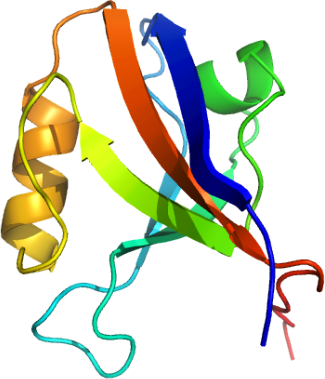
nMPDZA (10th domain)
PDB:2OPG
Revision
Revision Type:created
Revised by:created
Revision Date:created
Entry Clone Accession:MPDZA-s001
Entry Clone Source:Origene
SGC Clone Accession:Tag:Host:BL-21(DE3)R3 phage resistant
Construct
Prelude:Sequence:smGCETTIEISKGRTGLGLSIVGGSDTLL GAIIIHEVYEEGAACKDGRLWAGDQILEV NGIDLRKATHDEAINVLRQTPQRVRLTLY RDEAPYKstrl
Vector:pNIC28-Bsa4
Growth
Medium:LB
Antibiotics:Procedure: Transformed 90 µl competent BL-21 (DE3) phage resistant cells containing the Rosetta plasmid with 2 µl of the plasmid DNA and plated out onto LB plate plus 50 µg/ml kanamycin and 34 µg/ml chloramphenicol. The next day colonies were picked out into 40 mls of LB + 50 µg/ml kanamycin + 34 µg/ml chloramphenicol and grown at 37 degC overnight as a starter culture for a 1 litre growth. The large scale growth was grown at 37°C until approximately 30 mins before induction (OD600= 1.4) when the temperature was lowered to 22°C. After 1 hr 20 mins protein production was induced with the addition of 0.5 mM IPTG.
The next day cells were harvested by centrifugation at 6500 rpm for 15 minutes and resuspended in Lysis buffer before storage in the -80°C freezer.
Purification
ProcedureColumn 1: Ni-affinity, HisTrap, 1 ml (GE/Amersham)
Procedure: The cell extract was loaded on the column at 0.8 ml/min on an AKTA-express system (GE/Amersham). The column was then washed with 10 column volumes of Binding Buffer, 10 column volumes of Wash Buffer and the MPDZ domain eluted with Elution Buffer at 0.8 ml/min. The eluted peak of A280 was automatically collected.
Column 2 : Gel filtration, Hiload 16/60, S75 16/60 - 120 ml
Enzymatic treatment : At this stage the purity of the protein was greater than 95 % based on SDS-PAGE analysis. The C-terminal hexahistidine tag was removed by TEV protease treatment. The TEV protease, a hexahistidine-tagged construct, was over-expressed and purified in-house to a final concentration of 2.5 mg/ml.
Add 125 µl of the TEV protease was added to each fraction and left at 4°C overnight. The following steps were carried out to remove the cleaved products and TEV protease.
Place 200 µl of 50 % Ni-NTA agarose in a 1.5 ml eppendorf tubes, add 1ml of 50 mM Tris pH 8, 150 mM NaCl mix, spin down and remove buffer. Repeat this resin wash step once.
Add the TEV treated protein sample to the resin and mix for 60 min at 4°C . Finally spin down resin and collect the supernatant which contains the cleaved MPDZA domain. The supernatant was filtered through a 0.45µm syringe filter. The sample was then concentrated to 37.6 mg/ml using a 5 kD cutoff spin concentrator before storage in a -80°C freezer.
Extraction
ProcedurePMSF (final concentration 10 mM) in 10 ml of Lysis/ Binding Buffer and added to the thawed cell pellet (approximately 40mls volume) and mixed by inversion. The cell pellet was lysed by passing it four time through the EmulsiFlex C5 high pressure homogeniser, collecting a final volume of approximately 50mls. PEI was added to a final concentration of 0.15% and the cell debris and precipitated DNA were spun down at 16,500rpm for 45 mins). The supernatant was collected and manually filtered through a 0.22 µm filter.
Concentration:LigandMassSpec:Crystallization:Crystals grew from a 2:1 ratio mix of MPDZ 10th PDZ domain-to-reservoir (0.1 M Bis-Tris pH 6.5, 20% mPEG 5000) at 20°C. The crystals were cryoprotected by adding to the mother liquor ethylene glycol to a final concentration of 20%.
NMR Spectroscopy:Data Collection:Data were collected at the SLS X10SA beamline using a MAR CCD 225 detector. The wavelength was set to 0.9788A.
Data Processing:Data were analysed using MOSFLM and SCALA, while phases were calculated using PHASER and using as probes the homologues structures 2fne and 2awx. Two molecules were found in the asymmetric unit. The structure was refined using REFMAC.