Vector: pET24a. Details [PDF]; Sequence [FASTA] or [GenBank] |
Tag and addition: None |
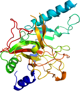
Protein sequence:
MACLSPSQLQKFQQDGFLVLEGFLSAEEC
VAMQQRIGEIVAEMDVPLHCRTEFSTQEE
EQLRAQGSTDYFLSSGDKIRFFFEKGVFD
EKGNFLVPPEKSINKIGHALHAHDPVFKS
ITHSFKVQTLARSLGLQMPVVVQSMYIFK
QPHFGGEVSPHQDASFLYTEPLGRVLGVW
IAVEDATLENGCLWFIPGSHTSGVSRRMV
RAPVGSAPGTSFLGSEPARDNSLFVPTPV
QRGALVLIHGEVVHKSKQNLSDRSRQAYT
FHLMEASGTTWSPENWLQPTAELPFPQLY
T |
Host: E.coli BL21(DE3) |
Cell Growth: Cells were grown at 370C in 2 x TY medium including 15 µg/ml kanamycin with shaking speed 200 rpm. Protein expression was induced by adding 0.5 mM IPTG at OD550=1.4. Cells were grown for another 4 hours and then harvested by centrifugation. |
Cell lysis: The cell pellet was resuspended in lysis buffer containing 50 mM HEPES, 10% glycerol, 2 mM DTT, 3 mM EDTA, pH 7.5. Lysozyme and DNase I were added to the cell suspension at 1 mg/ml and 20 µg/ml respectively, and the suspension was incubated at 40°C for 20 minutes. Cells were lysed by sonication followed by centrifugation to remove cell debris. The clarified supernatant was loaded onto the first column in the next step. |
Column 1: DEAE Sepharose fast flow 50/25 150 ml bedvolume ion-exchange column. The column was equilibrated with buffer A containing 50 mM HEPES, 10% glycerol, 2mM DTT, 2 mM EDTA and pH7.5. |
Procedure: The supernatant was loaded onto the column and washed with 3 column volume of buffer A. PHYD1 was eluted using a linear gradient from 0% to 30% buffer B (buffer A containing 1 M NaCl) in 6 column volumes. Fractions containing PHYD1 were pooled and concentrated. |
Column 2: Superdex-75 700 ml gel-filtration column. |
Procedure: The concentrated fractions containing the PHYD1 was loaded onto the gel filtration column and eluted with buffer C containing 25 mM Tris, 10% glycerol, 1mM DTT, 2 mM EDTA and pH 7.5. The fractions containing PHYD1 as judged by SDS-PAGE were pooled and subjected to a final step of purification. |
Column 3: Mono Q 16/10 ion-exchange column. |
Procedure: The pooled fractions were loaded directly to the Mono Q column pre-equilibrated with buffer D containing 25 mM Tris, 10% glycerol, 1 mM DTT, 1 mM EDTA and pH 8.0. PHYD1 was eluted with a gradient from 0 to 15% of buffer E (buffer D containing 1 M NaCl) in 5 column volumes. The fractions containing PHYD1 were pooled, concentrated, aliquoted and stored at -80°C for crystallographic and enzyme activity assay studies. |
Mass spectrometry: The ion-electronspray mass spectrometry measurement of the sample gives molecular weight of 32280 Da comparing to the calculated one of 32411 Da, suggesting that the n-terminal methionine is removed after translation. |
Crystallisation: Crystal screens were initially set up with sitting drop using 100 nl protein plus 100 nl well solution. Crystals were obtained with the screening condition of 0.2 M calcium acetate, 0.2 M potassium chloride 20% PEG 3350. Macro seeding is then used to obtain single and better crystals for structure studies. The Cryoprotectant used the mother solution with glycerol added to a final concentration 20% |
Data collection, Resolution: 1.9 Å, space group hexagonal P3(1)21, X-ray source: Synchrotron SLS-X10SA, single wavelength. |