RGS18A
PDB:2OWI
Revision
Revision Type:created
Revised by:created
Revision Date:created
Entry Clone Accession:IMAGE:4714909
Entry Clone Source:MGC
SGC Clone Accession:Tag:N-terminal, TEV cleavable (*) hexahistidine tag. Tag sequence: mhhhhhhssgvdlgtenlyfq(*)sm
Host:E. coli BL21(DE3)-Rosetta
Construct
Prelude:Sequence:smVSPEEAVKWGESFDKLLSHRDGLEAFT RFLKTEFSEENIEFWIACEDFKKSKGPQQ IHLKAKAIYEKFIQTDAPKEVNLDFHTKE VITNSITQPTLHSFDAAQSRVYQLMEQDS YTRFLKSDIYLDLMEGRPQRPTNLRRRSR SFTCNE
Vector:pLIC-SGC1
Growth
Medium:Antibiotics:Procedure:Expression testing: BL21(DE3)-Rosetta competent cells were transformed with the expression plasmid and plated on LB plates containing 60 µg/ml carbenicillin and 30µg/ml chloramphenicol. 4 Colonies from the transformation were used to inoculate 4 times 2 ml LB containing 60 µg/ml carbenicillin and 30µg/ml chloramphenicol. The cells were then grown at 700 RPM and 37°C for 6 hrs. The temperature was reduced to 22°C and the cells were induced with 1 mM IPTG. Expression of the 4 clones was analyzed by SDS - PAGE . The best clone was used for large scale expression in either [15N]-M9 or [13C, 15N]-M9 medium, containing 0.5g [15N]-NH4Cl per L and, for 13C-labelling, 2g [13C]-glucose per L.
Cell growth and induction: Starter overnight cultures of 80 ml were grown at 37°C in either [15N]-M9 or [13C,15N]-M9 supplemented with 60 µg/ml carbenicillin and 30µg/ml chloramphenicol. The large scale cultures were grown in 4 x 450 ml M9 with 60 µg/ml carbenicillin and 30µg/ml chloramphenicol in 4 x 2 L bottles (dilution for inoculation 1: 25). The culture was grown at 37°C and transferred to 22°C when the OD600 reached a value of 0.5. The culture was induced with 1 mM IPTG and grown at 22°C overnight. The next day the cells were harvested by centrifugation, washed with ice-cold 150 mM NaCl and frozen at -80°C.
Purification
ProcedureColumn 1 : Ni-affinity, MC-POROS, 8 ml (Applied Biosystems)
Procedure: The cell extract was loaded on the column at 1 ml/min on a workstation Vision (Applied Biosystems). The column was washed with 10 column volumes of start buffer and eluted with a gradient from 5 to 500 mM imidazole at a flow rate of 5 ml/min. The extinction at 280 nm was monitored and fractions were collected and analyzed by SDS - PAGE . Positive fractions were pooled and filled into a dialysis bag with 8 kDa MW cutoff.
TEV cleavage and dialysis: The His-tag was cleaved with 1 mg TEV per 40 mg target protein in a dialysis bag and dialysed with 5 L of: 20 mM Tris HCl 8.0, 500 mM NaCl, 1 mM ß mercaptoethanol, at 15°C overnight.
Column 2 : As column 1 but the flow rate was 0.5 ml/min and without imidazole in the start buffer.
Procedure: The flow through was collected and concentrated.
Concentration and buffer exchange: Using Amicon Ultra-15 concentrators with 5 kDa cutoff, all RGS 18-c008 samples were exchanged into NMR buffer and concentrated as follows:
- U-[15N]-labelled RGS 18A: final 1 mM protein in 20 mM phosphate buffer pH 6.0, 50 mM NaCl, 1 mM dDTT, 0.02% sodium azide.
- U-[13C,15N]- labelled RGS 18A: final 1.07 mM protein in 20 mM phosphate buffer pH 6.0, 50 mM NaCl, 1 mM dDTT, 0.02% sodium azide.
The latter was exchanged into D2O for acquisition of 13C-NOESY-HMQC, HCCH-TOCSY and HCCH-COSY experiments.
Concentrations were determined from the absorbance at 280 nm.
Extraction
ProcedureFrozen cell pellets from 0.9 L culture (ca. 2.5 g wet mass) were resuspended in a total volume of 25 ml lysis buffer. The cells were broken by 2 passes at 16,000 psi through a high pressure homogeniser followed by centrifugation for 20 minutes at 60,000g. The supernatant was further clarified by filtration (0.45 µm).
Concentration:LigandMassSpec:Calculated mass of the construct was 17760 Da (15N: 17977 Da / 13C,15N: 18768 Da). The determined mass for the U-[15N] sample was 18026 Da and for the U-[15N, 13C] sample 18106 Da. and 18791 Da (two main peaks out of 6)
Crystallization:NMR Spectroscopy:Data Collection:NMR spectra were acquired at 297 K, using Bruker DRX 600 and DMX 750 spectrometers in standard configuration with triple resonance probes equipped with self-shielded triple axis gradient coils. Spectra for the resonance and NOE assignment were recorded essentially as described in the original references. A 1 mM 15N-labelled RGS 18 sample in 90% H2O/10% D2O (NMR buffer; pH 6.0) was used for 3D 15N-separated NOESY-HSQC, 15N T1 and 15N T2 relaxation, and heteronuclear 15N- 1H NOE experiments. A 1 mM 13C, 15N-labelled sample of RGS 18 in 90% H2O/10% D2O (NMR buffer; pH 6.0) was used for all H N -detected triple resonance experiments, 3D CBCA(CO)NNH, CBCANNH, CC(CO)NNH, H(CCCO)NNH, HBHA(CBCACO)NNH, HNCO, HN(CA)CO, and for a 3D 13C-separated, aliphatic-centred NOESY-HSQC spectrum . The sample was then freeze-dried and redissolved in 100% D2O for acquisition of 3D 13C-separated HMQC-NOESY, HCCH-COSY, HCCH-TOCSY and 2D NOESY and TOCSY spectra. Data were processed using the program XWIN-NMR (version 2.6) of Bruker BioSpin GmbH ( Rheinstetten , Germany ).
Assignment: Assignment of 13C, 15N and 1H resonances was carried out using standard assignment procedures on Silicon Graphics O2 workstations and an Intel Dual Xeon 3GHz PC, with the interactive program CCPNMR Analysis version 1.0.9 ( Vranken et al., Proteins 59, 687-696; http://www.ccpn.ac.uk/ ).
The assignments are deposited in the BioMagResBank (http://www.bmrb.wisc.edu/) under accession code BMRB-7106.
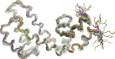
Data Processing:The three dimensional structure of RGS18A was calculated using the resonance assignments, the NOE peak lists from the 13C HMQC-NOESY, 13C NOESY-HSQC, 2D NOESY and 15N NOESY-HSQC spectra, and dihedral restraints predicted by TALOS (Cornilescu et al., J. Biomol. NMR, 13 (1999) 289-302; http://spin.niddk.nih.gov/NMRPipe/talos/) as input for the program CYANA v. 2.1 (Güntert et al., (1997) J. Mol. Biol. 273, 283-298; Herrmann et al., (2002). J. Mol. Biol. 319, 209-227). A tight ensemble of structures similar to those of other RGS domains was obtained, which were then refined manually. Hydrogen bond restraints were added on the basis of hydrogen exchange data and further NOEs were assigned based on the structures. Iterative refinement was carried out using XPLOR-NIH v. 2.14 (Schwieters et al., (2003) J. Magn. Reson. 160, 66-74; http://nmr.cit.nih.gov/xplor-nih/).
The refined ensemble of the 20 lowest energy structures was submitted to the PDB under code 2JM5.