DDX20
PDB:2OXC
Revision
Revision Type:created
Revised by:created
Revision Date:created
Entry Clone Accession:gi|23270929
Entry Clone Source:Mammalian Gene Collection
SGC Clone Accession:DDX20A-s001
Tag:N-terminal hexahistidine tag with integrated TEV protease cleavage site: mhhhhhhssgvdlgtenlyfq*sm
Host:E.coli BL21-Gold(DE3)pRARE2, where BL21-Gold(DE3) cells (Stratagene) have been transformed with pRARE2 originating from the Rosetta2 strain (Novagen). The pRARE2 plasmid supplies tRNAs for rare codons.
Construct
Prelude:Sequence:mhhhhhhssgvdlgtenlyfq*smRTAQDLSSPRTRTGDVLLAEPADFESLLLSRPVLEGLRAAGFERPSPVQLKAIPLGRCGLDLIVQAKSGTGKTCVFSTIALDSLVLENLSTQILILAPTREIAVQIHSVITAIGIKMEGLECHVFIGGTPLSQDKTRLKKCHIAVGSPGRIKQLIELDYLNPGSIRLFILDEADKLLEEGSFQEQINWIYSSLPASKQMLAVSATYPEFLANALTKYMRDPTFVRLNS
Vector:pNIC-Bsa4
Growth
Medium:Antibiotics:Procedure:Cells from a glycerol stock were grown in 20 ml TB supplemented with 8 g/l glycerol, 100 µg/ml kanamycin and 34 µg/ml chloramphenicol at 30 ºC overnight. The overnight culture (20 ml) was used to inoculate 1.5 l TB supplemented with 8 g/l glycerol, 50 µg/ml kanamycin and approximately 200 µl BREOX FMT 30 anti-foam solution (Cognis Performance Chemicals UK Ltd). The culture was grown in a LEX bioreactor system (Harbinger Biotechnology) at 37 ºC until OD600 reached ~1.7. The culture was down-tempered to 18 ºC over a period of 1 hour before target expression was induced by addition of 0.5 mM IPTG. Expression was allowed to continue overnight and cells were harvested the following morning by centrifugation (5,500 x
g, 10 min, 4 ºC). The resulting cell pellet (31.8 g wet cell weight) was resuspended in lysis buffer (1.5 ml/g cell pellet), supplemented with one tablet of Complete EDTA-free protease inhibitor (Roche Applied Science). The cell suspension was stored at -80 ºC.
Purification
ProcedureColumns
IMAC: Ni-charged 1 ml HiTrap Chelating HP (GE Healthcare)
Gel filtration column: HiLoad 16/60 Superdex 75 Prep Grade (GE Healthcare)
Procedure
Purification of the protein was performed as a two step process on an ÄKTAxpress system (GE Healthcare). Prior to purification, columns were equilibrated with IMAC wash1 buffer and gel filtration buffer, respectively. The filtered lysate was loaded onto the Ni-charged HiTrap Chelating column and washed with IMAC wash1 buffer followed by IMAC wash2 buffer. Bound protein was eluted from the IMAC column with IMAC elution buffer and automatically loaded onto the gel filtration column. Fractions containing the target protein were pooled and fresh TCEP was added to a final concentration of 2 mM.
Tag removal
The N-terminal histidine tag was proteolytically removed by incubating the target protein with His-tagged TEV protease at a molar ratio of 50:1 at 4 ºC overnight. The proteolytic reaction did not reach completion, as judged by SDS-PAGE. Target protein was purified from uncleaved protein, tag and protease by passing the reaction mixture over a Ni-charged 1 ml HisTrap FF column (GE Healthcare) pre-equilibrated with GF buffer (500 mM NaCl and 10 mM imidazole). The cleaved protein was concentrated and the buffer was exchanged to 20 mM HEPES, 500 mM NaCl, 10% glycerol, 2 mM TCEP, pH 7.5, using a Vivaspin 20 centrifugal filter device with 10,000 MWCO (Sartorius). The final protein concentration was determined to 33.8 mg/ml in a volume of 0.17 ml and the identity of the protein was confirmed by mass spectrometry.
Extraction
ProcedureThe cell suspension was quickly thawed in water and 2000 U Benzonase (Merck) were added. Cells were disrupted by sonication (Vibra-Cell, Sonics) at 80% amplitude for 3 min effective time (pulsed 4s on, 4s off) and cell debris was removed by centrifugation (49,100 x
g, 20 min, 4 ºC). The supernatant was decanted and filtered through a 0.45 µm flask filter.
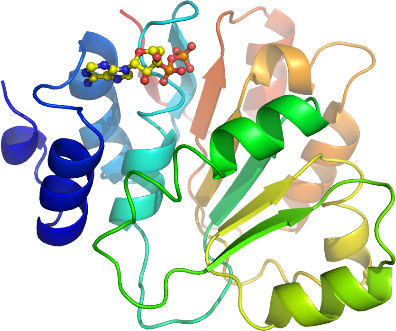
Concentration:LigandMassSpec:Crystallization:The crystal were obtained by the sitting drop vapour diffusion method in a 96-well plate. 0.1 µl protein solution (diluted to 20 mg/ml) including 20 mM ADP and 10 mM MgCl2 was mixed with 0.1 µl of well solution consisting of 0.1 M bis-Tris, pH 5.5, 0.2 M NaCl and 12% PEG 3350. The plate was incubated at 4 ºC and crystals appeared after one day and continued to grow for one more week. The crystal was briefly transferred to a cryo solution consisting of well solution complemented with 28% glycerol, and flash frozen in liquid nitrogen.
NMR Spectroscopy:Data Collection:Data to 1.3 Å resolution was collected at ESRF beamline ID14-2.
Data Processing:The structure was solved by molecular replacement with MOLREP using the N-terminal domain of eukaryotic translation initiation factor 4A (elF4A) from yeast as a search model (PDB entry: 1QVA). The asymmetric unit contained two protein monomers. The space group was P3121 with cell dimensions a=b=63.8 Å, c=214.3 Å. Refmac5 was used for refinement and Coot for model building. TLS restrained refinement using 4 TLS groups/monomer was used in the refinement process. The TLS groups were selected using the tlsmd server
http://skuld.bmsc.washington.edu/~tlsmd/. Data in the interval 38.5-1.3 Å resolution was used and at the end of the refinement the R values were: R= 15.4% and Rfree= 17.5%. A few residues in the N- and C-terminals were disordered and not visible in the electron density map. Coordinates for the crystal structure were deposited in the Protein Data Bank, accession code 2OXC.