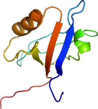
NHERF-1 (2nd PDZ domain): Human solute carrier family 9 isoform 3 regulator 1 - 2nd PDZ domain
PDB:2OZF
Revision
Revision Type:created
Revised by:created
Revision Date:created
Entry Clone Accession:NM_004252; gi|4759140
Entry Clone Source:Origene
SGC Clone Accession:SLC9A3R1A-c017
Tag:N-terminal hexahistidine tag followed with a TEV cleavage site. C-terminal PDZ binding motif (ETSL).
Host:BL-21(DE3)R3 phage resistant
Construct
Prelude:C-terminal PDZ binding motif ( ETSL )
Sequence:mhhhhhhssgvdlgtenlyfq*smLRPRL CTMKKGPSGYGFNLHSDKSKPGQFIRSVD PDSPAEASGLRAQDRIVEVNGVCMEGKQH GDVVSAIRAGGDETKLLVVDRETDEFetsl
Vector:pNIC28-Bsa4
Growth
Medium:TB
Antibiotics:Procedure:A glycerol stock was used to innoculate a starter culture of 50 ml LB with 50 microG/ml kanamycin. The next day 15 mL of the starter culture was used to innoculate 1 litre of TB containing 50 microG/ml kanamycin. The culture was grown at 37degC overnight as a starter culture for a 1 litre growth. The large scale growth was grown at 37degC until an OD600 of 4.2 was reached when the temperature was lowered to 25degC. One hour later protein production was induced with the addition of 0.5 mM IPTG and the cells cultured overnight.
The next day cells were harvested by centrifugation at 6500 rpm for 15 minutes and resuspended in Lysis buffer (Lysis Buffer: 50 mM HEPES pH 7.5, 500 mM NaCl, 5 % Glycerol, 5 mM Imidazole pH 7.5, 0.5 mM TCEP)before storage in the -80°C freezer.
Purification
Procedure Ni-affinity: The cell extract (supernatant) was loaded on the column 1.5ml bed volume of Ni-NTA (Qiagen). The column was then washed with 15 column volumes of Binding Buffer to equilibriate the resin, 40 column volumes of Binding Buffer, 20 column volumes of Wash Buffer and the SLC9A3R1A protein eluted with 10 column volumes of Elution Buffer and collected in 2ml fractions.
Enzymatic treatment : At this stage the purity of the protein was greater than 95 % based on SDS-PAGE analysis. The C-terminal hexahistidine tag was removed by TEV protease treatment. The TEV protease, a hexahistidine-tagged construct, was over-expressed and purified in-house to a final concentration of 2.5 mg/ml.
Add 250 µl of the TEV protease was added to the pooled fractions and left at 4degC overnight. The following steps were carried out to remove the cleaved products and TEV protease.
Place 200 microL of 50 % Ni-NTA agarose in a 1.5 ml gravity column, add 1ml of 50mM HEPES pH 7.4, 500mM NaCl, 0.5mM TCEP to wash the resin (GF buffer). Resuspend in GF buffer and add the TEV treated protein sample to the resin for batch binding over 60 min at 4degC. Finally reform the column and collect the supernatant which contains the cleaved SYN3A domain. The supernatant was filtered through a 0.45micrometer syringe filter.
Gel filtration: The TEV treated sample was loaded on the gel filtration column in GF buffer at 1.0 ml/min. Eluted proteins were collected in 1 ml fractions. Mass spec analysis indicated that the sample was not fully cleaved hence fractions containing SLC9A3R1A were pooled and treated overnight with 200 µl of TEV protease at 4°C . Complete tag cleavage was determined by Mass Spec before concentration of the fractions into a final volume of 2 ml. Next the sample was filtered with a 0.2 micron Millipore syringe filter.
Column 3: 1.5ml Ni-affinity Qiagen gravity column . Gel filtration: To remove any remaining contaminants, the cleaved products and TEV protease were removed by Ni-NTA batch binding as previously described with 1.5ml Ni-affinity Qiagen for 1 hour before collecting the eluatant (cleaved pure protein) which passes through the gravity column.
Concentration: The sample was then concentrated to 99 mg/ml using a 5 kD MW cutoff spin concentrator before storage in a -80°C freezer.
Extraction
ProcedurePMSF (final concentration 10 mM) in 10 ml of Lysis/Binding Buffer and added to the thawed cell pellet (approximately 40 mL volume) and mixed by inversion. The cell pellet was lysed by passing it four times through the EmulsiFlex C5 high pressure homogeniser, collecting a final volume of approximately 50 mls. PEI was added to a final concentration of 0.15% mixed by inversion and the cell debris and precipitated DNA were spun down at 21,500rpm for 45 mins).
Concentration:LigandMassSpec:Expected MWt: 10320.5; Measured MWt: 10302.4
Crystallization:Crystals grew from a 1:1 ratio mix of SLC9A3R1A-to-reservoir (0.20M NaNO 3 ; 0.1M BTprop; 20% PEG 3350; 10% ethylene glycol pH 8.5) at 20°C. Crystal form was plates. Cryoprotection 20% Ethylene glycol.
NMR Spectroscopy:Data Collection:Data Processing: