HSD17B8
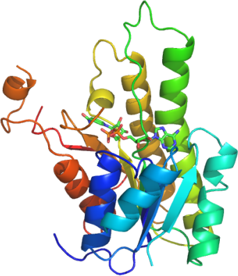
PDB:2PD6
Revision
Revision Type:created
Revised by:created
Revision Date:created
Entry Clone Accession:gi|15277342
Entry Clone Source:HSD17B8A-c027
SGC Clone Accession:
Tag:shhhhhhdykddddk
Host:BL21(DE3)-R3-pRARE2
Construct
Prelude:
Sequence:mQNRLRSALALVTGAGSGIGRAVSVRLAGEGATVAACDLDRAAAQETVRLLGGPGSKEGPPRGNHAAFQADVSEARAARCLLEQVQACFSRPPSVVVSCAGITQDEFLLHMSEDDWDKVIAVNLKGTFLVTQAAAQALVSNGCRGSIINISSIVGKVGNVGQTNYAASKAGVIGLTQTAARELGRHGIRCNSVLPGFIATPMTQKVPQKVVDKITEMIPMGHLGDPEDVADVVAFLASEDSGYITGTSVEVTGGLFMAENLYFQ*shhhhhhdykddddk
Vector:pNIC-CTHF
Growth
Medium:
Antibiotics:
Procedure:A glycerol stock grown from a single colony in TB (+ 50 mg/ml kanamycin) was used to inoculate 10 ml of TB supplemented with 50 mg/ml kanamycin. This starter culture was grown overnight at 37 degC and used to inoculate a 1 liter culture in the same medium. The culture was grown at 37 degC until the OD600 reached ~2.0. After that the temperature was lowered to 18 degC. Protein production was induced with 1mM IPTG and recombinant HSD17B8A was expressed at that temperature over night. The next day cells were harvested by centrifugation at 4000 rpm for 15 minutes. The cell pellet was stored at -80 degC.
Purification
Procedure
All purification steps were carried out using an AKTAexpress system (GE Healthcare) at 7ºC. The lysate was loaded on a pre-equilibrated His-trap column at 0.8 ml/min. After loading, the column was washed at 0.8 ml/min with 10 ml binding buffer, then 20 ml wash buffer, and protein was eluted with 5 ml of elution buffer. The peak fraction was collected automatically according to A280. The C-terminal 6xHis-tag was cleaved by incubating the protein overnight with TEV protease.
The HSD17B8A containing fraction eluted of the Ni-affinity chromatography was automatically loaded on the SEC column at 1.2 ml/min. Eluted fractions were 95% pure as judged by SDS-PAGE.
Extraction
Procedure
The cell pellet (55 g) was re-suspended in one volume (55 ml) of 2x extraction buffer. The re-suspended cells were lysed by one passage through a Constant Systems cell breaker and subsequent sonication. DNA was precipitated by addition of PEI to a final concentration of 0.15 % during an incubation time of 30 min on ice, followed by a centrifugation at 17,000 rpm (4 degC); The supernatant was further cleared by filtration through a 0.2 microm serum Acrodisc filter.
Concentration:13 mg/ml in SEC buffer using a c entricon with a 10kDa cut off
Ligand
MassSpec:ESI-MS revealed that the protein had the expected mass of 27440 Da.
Crystallization:Crystals were grown at 20 degC in 150nl sitting drops using a 1:1 mix of HSD17B8A (13mg ml-1) plus 5mM NAD+, with a solution containing 200 mM sodium fluoride, 20% PEG3350 and 10% ethylene glycol. The crystal used for data collection was cryoprotected using 20% ethylene glycol.
NMR Spectroscopy:
Data Collection:Resolution: 2.00 Å; X-ray source: Beamline SLS -X10SA, single wavelength (λ= 1.00234).
Data Processing: