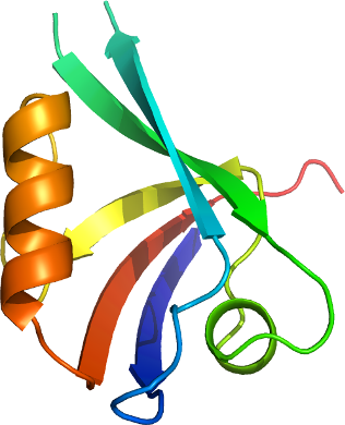
GRASP
PDB:2PNT
Revision
Revision Type:created
Revised by:created
Revision Date:created
Entry Clone Accession:gi|32171221
Entry Clone Source:MGC
SGC Clone Accession:GRASPA-c008
Tag:N-terminal, TEV cleavable hexahistidine tag
Host:BL21(DE3)-R3-pRARE2 (A homemade phage resistant version of BL21(DE3) containing the pRARE2 plasmid from Rosetta II (DE3) cells).
Construct
Prelude:
Sequence:smQQRKVLTLEKEDNQTFGFEIQTYGLHHREEQRVEMVTFVCRVHESSPAQLAGLTPGDTIASVNGLNVEGIRHREIVDIIKASGNVLRLETLYSSTL
Vector:pNIC28-Bsa4.
Growth
Medium:
Antibiotics:
Procedure:The construct DNA was transformed into homemade chemically competent cells of the expression strain by a standard heat shock procedure. A number of colonies from the transformation were used to innoculate 1 ml of LB media containing 50 µg/ml kanamycin and 34 µg/ml chloramphenicol, which was placed in a 37°C shaker overnight. The next day glycerol stocks were prepared from this overnight culture.
A glycerol stock was used to innoculate 40 ml of LB media containing 50 µg/ml kanamycin and 34 µg/ml chloramphenicol, which was placed in a 37°C shaker overnight. The next day this starter culture was used to innoculate 2x 1L of TB media (18 ml starter culture into each) containing 50 µg/ml kanamycin. After 5 hours the temperature was reduced to 18°C. After a further 1.5 hours the cells were induced by the addition of 1.0 mM IPTG. The expression was continued overnight.
Cells were spun at 6000 rpm, JLA8.1000 rotor, for 15 mins at 4°C. The cell pellets were placed in a -80°C freezer
Purification
Procedure
Column 1: HisTrap 1ml.
Column 2: Gel filtration. Hiload S200 16/60 - 120 ml volume.
The protein was purified using an AktaExpress system.
The clarified cell extract was passed through the column 1 at a flow rate of 0.8 ml/min. The column was then washed Binding Buffer until a stable UV baseline was achieved. The column was then washed with Wash Buffer until a stable UV baseline was achieved. The protein was eluted with 5 ml of Elution Buffer.
The column 2 was pre-equilibrated with Gel Filtration Buffer. The HisTrap eluant was loaded on the gel filtration column automatically after the HisTrap elution at a flow rate of 1.2 ml/min. Eluted proteins were collected in 1.8 ml fractions. The fractions containing protein were identified on a coomasie blue stained gel.
TEV protease digestion: The gel filtration fractions containing GRASPA were pooled and 170 µl of TEV protease solution (about 1 mg/ml) was added. The digestion was left overnight at 4°C.Rebinding of impurities to Ni-NTA: The protein was mixed with Ni-NTA resin (0.25 ml, pre-equilibrated into Gel Filtration Buffer) at 4°C for 60 minutes. The resin was spun down and the supernatent collected.
Extraction
Procedure
The cell pellet was resuspended in 50 ml of lysis buffer containing 0.5 mM PMSF. The resuspended cell pellet was passed 4 times through an Emulsiflex C5 high-pressure homogeniser, collecting a final volume of approximately 200 ml after dilution with Lysis Buffer. PEI was added to a final concentration of 0.2 % and the cell debris and precipitated DNA were spun down (17500 rpm, JA18 rotor, 90 min). The supernatent was filtered through a 0.2 m M syringe filter.
Concentration:The TEV protease cleaved GRASPA was concentrated to 50 mg/ml (measured using a nanodrop machine), distributed into aliquots and frozen at -80°C.
Ligand
MassSpec:Measured: 11038.3; Expected: 11038.4
Crystallization:Crystals grew from a 2:1 ratio of protein to precipitant solution (1.26 M NaH2PO 4 , 0.14 M K 2 HPO 4 ), using the vapour diffusion method.
NMR Spectroscopy:
Data Collection:Crystals were cryo-protected by equilibration into precipitant solution containing 30% glycerol, and then flash frozen in liquid nitrogen. Data was collected to a resolution of 2.15 Å at the Swiss Light Source, beamline X10SA.
Data Processing: