IDI2
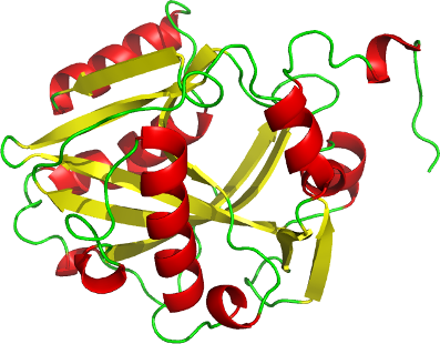
PDB:2PNY
Revision
Revision Type:created
Revised by:created
Revision Date:created
Entry Clone Accession:NP_150286
Entry Clone Source:OBS-BC017778.1
SGC Clone Accession:idi2.001.227:45A03
Tag:mgsshhhhhhssglvprgs
Host:BL21(DE3)
Construct
Prelude:
Sequence:mgsshhhhhhssglvprgsMSDINLDWVDRRQLQRLEEMLIVVDENDKVIGADTKRNCHLNENIEKGLLHRAFSVVLFNTKNRILIQQRSDTKVTFPGYFTDSCSSHPLYNPAELEEKDAIGVRRAAQRRLQAELGIPGEQISPEDIVFMTIYHHKAKSDRIWGEHEICYLLLVRKNVTLNPDPSETKSILYLSQEELWELLEREARGEVKVTPWLRTIAERFLYRWWPHLDDVTPFVELHKIHRV
Vector:p28a-thrombin-lic
Growth
Medium:Terrific Broth (TB)
Antibiotics:
Procedure:Idi2 was expressed in E. coli BL21 (DE3) grown in Terrific Broth (TB) in the presence of 50 µg/ml of kanamycin at 37ºC to an OD600 of 7.5. Cells were then induced by isopropyl-1-thio-D-galactopyranoside (IPTG), final concentration 0.1 mM, and incubated overnight at 15ºC. The culture was centrifuged and the cell pellets were collected and stored at -80ºC.
Purification
Procedure
Column 1: His- Ni-NTA resin (Qiagen 30450)
Column 2: XK 16x65 column (GE Healthcare)
Lysed cells were diluted to 50 -100 mL final volume and imidazole added to a final concentration of 10 mM; this was then mixed with 2-3 mL of HisLink Protein Purification Resin (Promega V8821) per construct. The mixture was incubated with mixing for at least 20 minutes at 4 degC. The lysate was spun at 500xg for 3 minutes to pellet the HisLink resin. The lysate was carefully decanted off the resin, and then 50 mL of lysis buffer were added to wash the resin. The resin was allowed to settle for 5 minutes, then poured off and washed 3 more times with fresh lysis buffer. The washed resin was then loaded onto a gravity column, and then washed with a column volume of low imidazole buffer. Samples were eluted from the HisLink resin by exposure to 10-14 mL MAC elution buffer at a 1mL/min flow rate. An XK 16x65 column (part numbers 18-1031-47 and 18-6488-01, GE Healthcare) packed with HighLoad Superdex 200 resin (10-1043-04, GE Healthcare) was pre-equilibrated with gel filtration buffer for 1.5 column volumes at a flow rate of 1.5 mL/min. 10 mL of sample was loaded onto the column at 1.5 mL/min, and 2mL fractions were collected. Peak fractions were analyzed for purity and pooled. A mixture of MgCl2, MnCl2, and CaCl2 (final concentration of each: 1mM) was added to purified protein, and then the mixture was concentrated using 15 mL concentrators with 10,000 MWCO (Amicon, UFC901024) to a final concentration of 10 mg/mL for crystallographic screening.
Extraction
Procedure
Cell pellets contained in bags (Beckman 369256) obtained from 4L culture were thawed by soaking in warm water. Each cell pellet was resuspended in 20 mL lysis buffer and then homogenized using an Ultra-Turrax T8 homogenizer (IKA Works) at maximal setting for 30-60 seconds per pellet. Cell lysis was accomplished by sonication (Virtis408912, Virsonic) on ice: the sonication protocol is 10 sec pulse at half-maximal frequency (5.0), 10 second rest, for 6 minutes total sonication time per pellet.
Concentration:10 mg/mL
Ligand
MassSpec:
Crystallization:Diffraction quality crystals were grown using the following protocol: 2M sodium/potassium phosphate, pH 7.0 were mixed in a ratio of 0.5 microL protein+0.5 microL reservoir in sitting drop trays and placed at 18 degC. Resultant crystals were cryoprotected in 20% glycerol and frozen in liquid nitrogen.
NMR Spectroscopy:
Data Collection:
Data Processing: