PARP12
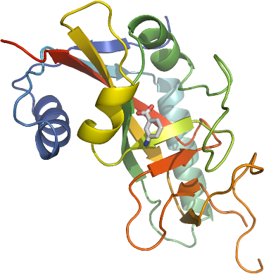
PDB:2PQF
Revision
Revision Type:created
Revised by:created
Revision Date:created
Entry Clone Accession:gi|51980617
Entry Clone Source:Mammalian Gene Collection
SGC Clone Accession:Tag:N-terminal hexahistidine tag with integrated TEV protease cleavage site: mhhhhhhssgvdlgtenlyfq*sm
Host:E.coli BL21(DE3) (Novagen)
Construct
Prelude:Sequence:mhhhhhhssgvdlgtenlyfq*smDSSALPDPGFQKITLSSSSEEYQKVWNLFNRTLPFYFVQKIERVQNLALWEVYQWQKGQMQKQNGGKAVDERQLFHGTSAIFVDAICQQNFDWRVCGVHGTSYGKGSYFARDAAYSHHYSKSDTQTHTMFLARVLVGEFVRGNASFVRPPAKEGWSNAFYDSCVNSVSDPSIFVIFEKHQVYPEYVIQYTTSSKPS
Vector:pNIC-Bsa4
Growth
Medium:Selenomethionine labeled protein was grown in minimal medium containing: 25 mM Na
2HPO
4, 25 mM KH
2PO
4, 50 mM NH
4Cl, 5 mM Na
2SO
4, 0.4% (w/v) glucose, 2 mM MgSO
4, 0.1 mM CaCl
2, 1.0 µM MnCl
2, 10 µM FeSO
4.
Mix of amino acids added per liter culture (Van Duyne, G. D.,
J. Mol. Biol. 229, 105-124 (1993)): 100 mg each of lysine, threonine, phenylalanine and 50 mg each of leucine, isoleucine, valine, L(+)-selenomethionine.
Antibiotics:Procedure:Cells from a glycerol stock were streaked onto LB-agar plates. 5-10 colonies were used to inoculate 2 x 40 ml LB supplemented with 100 µg/ml kanamycin and the cells were grown at 30 ºC overnight. 60 ml of the overnight culture were used to inoculate 6 bottles with 1.5 l minimal medium (without amino acids) supplemented with 50 µg/ml kanamycin and approximately 200 µl BREOX FMT 30 anti-foam solution (Cognis Performance Chemicals UK Ltd) per bottle. The cultures were grown in a LEX bioreactor system (Harbinger Biotechnology) at 37 ºC until OD600 reached ~0.6. The bottles were down-tempered to 18 ºC and after 10 minutes, amino acids were added. About one hour later, expression of target protein was induced by addition of 0.5 mM IPTG. The expression was allowed to continue at 18 ºC overnight. Cells were harvested the following morning by centrifugation (5,500 x
g, 15 min, 4 ºC). The resulting cell pellet (26.7 g from 9 liter culture) was resuspended in lysis buffer (2 ml/g cell pellet) supplemented with 0.5 tablet of Complete EDTA-free protease inhibitor (Roche Applied Science) and stored at -80 ºC.
Purification
ProcedureColumns
IMAC1: Ni-charged 1 ml HisTrap FF Crude (GE Healthcare)
IMAC2: Ni-charged 1 ml HiTrap Chelating HP (GE Healthcare)
Gel filtration column: HiLoad 16/60 Superdex 75 Prep Grade (GE Healthcare)
Procedure
Purification of the protein was performed as a two step process on an ÄKTAprime system (GE Healthcare). Prior to purification, columns were equilibrated with IMAC wash buffer and gel filtration buffer, respectively. The filtered lysate was loaded onto the Ni-charged HisTrap FF Crude column and washed with IMAC wash buffer. Bound protein was eluted from the IMAC column by a step wise gradient with IMAC elution buffer. Fractions containing the target protein were pooled and 2 mM TCEP was addded. The sample was concentrated to 3 ml before loaded onto the gel filtration column. The purified protein was subsequently concentrated using an Amicon Ultra-15 centrifugal filter device, 10,000 NMWL (Millipore) to 4.5 mg/ml in a volume of 1.5 ml. The identity of the protein was confirmed by mass spectrometry.
Tag removal
The N-terminal histidine tag was proteolytically removed by incubating the target protein with His-tagged TEV protease at a molar ratio of 30:1 at 4 ºC overnight. The proteolytic reaction went to completion, as judged by SDS-PAGE. Target protein was diluted in IMAC wash buffer and subsequently purified from tag and protease by passing the reaction mixture over a Ni-charged 1 ml HiTrap Chelating HP column (GE Healthcare) pre-equilibrated with IMAC wash buffer. The protein was concentrated and the buffer was changed to GF buffer (500 mM NaCl and 2 mM TCEP) using a Vivaspin 20 centrifugal filter device with 10,000 MWCO (Sartorius). The final protein concentration was determined to 23.5 mg/ml in a volume of 0.12 ml.
Extraction
ProcedureThe cell suspension was quickly thawed in water and 1500 U Benzonase (Merck) was added. Cells were disrupted by sonication (Vibra-Cell, Sonics) at 80% amplitude for 3 min effective time (pulsed 4s on, 4s off) and cell debris was removed by centrifugation (49,000 x
g, 30 min, 4 ºC). The supernatant was decanted and filtered through a 0.45 µm flask filter.
Concentration:LigandMassSpec:Crystallization:Crystals were obtained by the hanging drop vapour diffusion method in a 24-well plate containing 500 µl well solution. Before crystallization, 10 mM 3-aminobenzoic acid was added to the protein. 0.7 µl of the protein solution (diluted to 19 mg/ml) was mixed with 0.7 µl of well solution consisting of 0.1 M Na-citrate pH 4.3, 9% PEG 6000 and 2% DMSO. The plate was incubated at 20 ºC. Crystals appeared after one day and continued to grow for one more week to reach their maximal size (approx. 1000 µm × 50 µm × 50 µm). The crystals were quickly transferred to cryo solution containing well solution and 30% glycerol and flash frozen in liquid nitrogen.
NMR Spectroscopy:Data Collection:Diffraction data was collected at beamline BL 14-1 at the BESSY.
Data Processing:Data was indexed and integrated in space group I4 with the XDS package. The structure was solved by SAD Â Single Anomalous Dispersion using selenomethionine-labeled protein with the program SOLVE. The asymmetric unit contains six protein monomers. The cell dimensions are a=b=206.59 Å c=84.73 Å. Refmac5 was used for refinement and Coot for model building. TLS restrained refinement using 18 TLS groups was used in the refinement process. The TLS groups were selected using the tlsmd server
http://skuld.bmsc.washington.edu/~tlsmd/. Data in the interval 19.6-2.20 Å resolution was used and at the end of the refinement the R values were: R= 19.9% and Rfree= 24.5%. Coordinates for the crystal structure were deposited in the Protein Data Bank, accession code 2PQF.