GART
PDB:2QK4
Revision
Revision Type:created
Revised by:created
Revision Date:created
Entry Clone Accession:gi|4503915
Entry Clone Source:Mammalian Gene Collection
SGC Clone Accession:Tag:N-terminal hexahistidine tag with integrated TEV protease cleavage site: mhhhhhhssgvdlgtenlyfq*s(m).
Host:E.coli BL21-Gold(DE3)pRARE2, where BL21-Gold(DE3) cells (Stratagene) have been transformed with pRARE2 originating from the Rosetta2 strain (Novagen). The pRARE2 plasmid supplies tRNAs for rare codons.
Construct
Prelude:Sequence:mhhhhhhssgvdlgtenlyfq*smAARVLIIGSGGREHTLAWKLAQSHHVKQVLVAPGNAGTACSEKISNTAISISDHTALAQFCKEKKIEFVVVGPEAPLAAGIVGNLRSAGVQCFGPTAEAAQLESSKRFAKEFMDRHGIPTAQWKAFTKPEEACSFILSADFPALVVKASGLAAGKGVIVAKSKEEACKAVQEIMQEKAFGAAGETIVIEELLDGEEVSCLCFTDGKTVAPMPPAQDHKRLLEGDGGPNTGGMGAYCPAPQVSNDLLLKIKDTVLQRTVDGMQQEGTPYTGILYAGIMLTKNGPKVLEFNCRFGDPECQVILPLLKSDLYEVIQSTLDGLLCTSLPVWLENHTALTVVMASKGYPGDYTKGVEITGFPEAQALGLEVFHAGTALKNGKVVTHGGRVLAVTAIRENLISALEEAKKGLAAIKFEGAIYRKDIGFRAIAFLQ
Vector:pNIC-Bsa4
Growth
Medium:Antibiotics:Procedure:Cells from a glycerol stock were grown in 150 ml LB supplemented with 8 g/l glycerol, 50 µg/ml kanamycin and 34 µg/ml chloramphenicol at 37 ºC overnight. The overnight culture (120 ml) was used to inoculate 4 x 750 ml TB supplemented with 8 g/l glycerol, 50 µg/ml kanamycin and 34 µg/ml chloramphenicol. The cultures were grown in TunAir flasks at 37 ºC until OD600 reached ~1.5. The cultures were down-tempered to 18 ºC over a period of 1 hour before target expression was induced by addition of 0.5 mM IPTG. Expression was allowed to continue overnight and cells were harvested the following morning by centrifugation (5,500 x
g, 20 min, 4 ºC). The resulting cell pellet (93.8 g wet cell weight) was stored at -80 ºC.
Purification
ProcedureColumns
IMAC: Ni-charged 5 ml HiTrap Chelating HP (GE Healthcare)
Gel filtration column: HiLoad 16/60 Superdex 200 Prep Grade (GE Healthcare)
Procedure
Purification of the protein was performed as a two step process on an ÄKTAprime system (GE Healthcare). Prior to purification, columns were equilibrated with IMAC wash1 buffer and gel filtration buffer, respectively. The filtered lysate was loaded onto the Ni-charged HiTrap Chelating column and washed with IMAC wash1 buffer followed by IMAC wash2 buffer. Bound protein was eluted from the IMAC column with IMAC elution buffer and loaded onto the gel filtration column. Fractions containing the target protein were pooled and the protein was subsequently concentrated using a Vivaspin 6 centrifugal filter device, 10,000 MWCO (Sartorius) to 21.5 mg/ml in a volume of 0.72 ml. The identity of the protein was confirmed by mass spectrometry.
Extraction
ProcedureThe cell pellet was quickly thawed and resuspended in lysis buffer (0.5 ml/g cell pellet), supplemented with 1000 U Benzonase (Merck), one tablet of Complete EDTA-free protease inhibitor (Roche Applied Science) and a knife edge of lysozyme (Sigma). Cells were disrupted by sonication (Vibra-Cell, Sonics) at 80% amplitude for 3 min effective time (pulsed 4s on, 4s off) and cell debris was removed by centrifugation (49,000 x
g, 60 min, 4 ºC). The supernatant was decanted and filtered through a 0.45 µm syringe filter.
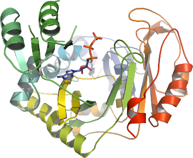
Concentration:LigandMassSpec:Crystallization:Crystals were obtained by the hanging drop vapour diffusion method in a 24-well plate containing 500 µl well solution. 1 µl of the protein solution (diluted to 15.7 mg/ml) including 4 mM ATP and 4 mM glycine, was mixed with 1 µl of well solution consisting of 0.1 M bis-Tris pH 5.2, 0.3 M Li sulfate and 16% PEG 3350. The plate was incubated at 20 ºC. The crystals were quickly transferred to cryo solution containing 0.1 M bis-Tris pH 5.2, 0.3 M Li sulfate and 17% PEG 3350, 0.2 M NaCl and 20% glycerol, and flash frozen in liquid nitrogen.
NMR Spectroscopy:Data Collection:Data was collected at ESRF beamline ID14-2.
Data Processing:Data was processed with XDS in space group P21 (a=70.28 Å, b=79.81 Å, c=212.72 Å and β=104.07Å). The structure was solved by molecular replacement using PDB entry 1GSO as a model after editing it with MrBUMP (CHAINSAW). The edited model was used as an input for MOLREP, which located the two molecules in the asymmetric unit. Model building was done with COOT and refinement with REFMAC5.