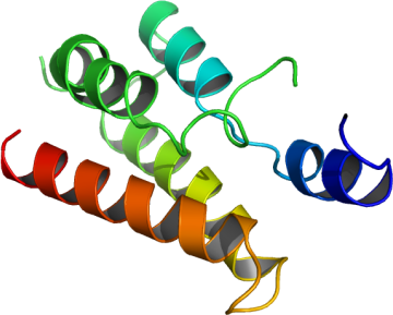
BRDT
PDB:2RFJ
Revision
Revision Type:created
Revised by:created
Revision Date:created
Entry Clone Accession:gi|46399198
Entry Clone Source:IMAGE
SGC Clone Accession:IMAGE:5742670
Tag:mhhhhhhssgvdlgtenlyfq*s(m) TEV-cleavable (*) N-terminal his6 tag.
Host:BL21(DE3)-R3-pRARE2
Construct
Prelude:
Sequence:mhhhhhhssgvdlgtenlyfq*smNTKKNGRLTNQLQYLQKVVLKDLWKHSFSWPFQRPVDAVKLQLPDYYTIIKNPMDLNTIKKRLENKYYAKASECIEDFNTMFSNCYLYNKPGDDIVLMAQALEKLFMQKLSQMPQEE
Vector: pNIC28-Bsa4.
Growth
Medium:
Antibiotics:
Procedure:1ml from a 10 ml overnight culture containing 50 µg/ml kanamycin was used to inoculate 1 liter of TB media containing 50 µg/ml kanamycin. Cultures were grown at 37°C until the OD600 reached ~2.0. After that the temperature was adjusted to 18°C. Expression was induced overnight using 1mM IPTG. The cells were collected by centrifugation and the pellets were frozen.
Purification
Procedure
Column 1: Ni-affinity chromatography ( HisTrap FF crude, 5 ml )All purification steps were carried out using an AKTAexpress system (GE Healthcare) at 7ºC. The lysate was loaded on a pre-equilibrated His-trap column at 0.8 ml/min, using a standard purification method. After loading, the column was washed at 0.8 ml/min with 10 ml binding buffer, then 20 ml wash buffer, and protein was eluted with 5 ml of elution buffer. The peak fraction was collected automatically according to A280.Column 2: Size exclusion chromatography (HiLoad 16/60 Superdex S75)The BRDTA containing fraction eluted of the Ni-affinity chromatography was automatically loaded on the SEC column at 1.2 ml/min. BRDTA eluted at a retention time corresponding to the monomeric protein. Eluted fractions were 95% pure as judged by SDS-PAGE , and confirmed by mass spectrometer (expected mass: 16614 Da).Tag cleavage and column 3: The protein was incubated with TEV protease overnight at 4ºC. After cleavage, the protein was passed through a Ni-sepharose column (0.5 ml bed volume) to capture the cleaved tag and other contaminants. The molecular mass after tag cleavage was confirmed by mass spectrometry (14149 Da).
Extraction
Procedure
The cell pellet (35 g) from 2 L cultue was re-suspended in one volume (35 ml) of 2x extraction buffer. The re-suspended cells were lysed by one passage through a high-pressure cell breaker (Constant Systems) and subsequent sonication; the cell breaker was washed with 1x extraction buffer, bringing the total volume to 120 ml. DNA was precipitation by addition of PEI (polyethyleneimine, pH 7.5) to a final concentration of 0.15 % during an incubation time of 30 min on ice, followed by a centrifugation at 17,000 rpm (4°C); The supernatant was further cleared by filtration through a 0.2 µm filter (serum Acrodisc).
Concentration:The protein was concentrated to 35 mg/ml using a Centricon device (5 kDa cut off).
Ligand
MassSpec:
Crystallization:Crystals were obtained using the vapor diffusion method. Drops were setup in 96 well sitting drop plates by mixing 100nl of the concentrated protein (35 mg/ml) with 100nl of a well solution containing 0.20M Na/K(tart); 20.0% PEG 3350; 10.0% EtGly.
NMR Spectroscopy:
Data Collection:Diffraction data were collected at 2.0 Å resolution from a crystal that was cryo-protected using 20% ethylene glycol (end concentration) at the SLS beam-line SAX10.
Data Processing: