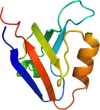
PDLIM5
PDB:2UZC
Revision
Revision Type:created
Revised by:created
Revision Date:created
Entry Clone Accession:gi|58533153
Entry Clone Source:MGC
SGC Clone Accession:PDLIM5A-c003
Tag:N-terminal, TEV cleavable hexahistidine tag
Host:BL-21(DE3)-R3-Rosetta (A homemade phage resistant version of BL21(DE3) containing the pRARE2 plasmid from Rosetta II (DE3) cells).
Construct
Prelude:
Sequence:smSNYSVSLVGPAPWGFRLQGGKDFNMPLTISSLKDGGKAAQANVRIGDVVLSIDGINAQGMTHLEAQNKIKGCTGSLNMTLQRESDL
Vector:pNIC28-Bsa4.
Growth
Medium:
Antibiotics:
Procedure:The construct DNA was transformed into homemade chemically competent cells of the expression strain by a standard heat shock procedure.
50 µl of a thawed glycerol stock was used to innoculate 40 ml of TB media containing 50 µg/ml kanamycin and 34 µg/ml chloramphenicol, which was placed in a 37°C shaker overnight. The next day this starter culture was used to innoculate 2x 1L of TB media (18 ml starter culture into each) containing 33 µg/ml kanamycin. When the OD600 reached ~2 the temperature was reduced to 20°C and the cells were induced by the addition of 1.0 mM IPTG. The expression was continued overnight (~ 14 hours).
Cells were spun at 6238x g for 7 mins at 4°C. Each 1L pellet was resuspended in 35 ml Resuspension Buffer. The resuspended cell pellets were placed in a -80°C freezer.
Purification
Procedure
Column 1: HisTrap 1ml.
Column 2: Gel filtration. Hiload S200 16/60 -120 ml volume.
The clarified cell extract was passed through the column 1 at a flow rate of 0.8 ml/min. The column was then washed Binding Buffer until a stable UV baseline was achieved. The column was then washed with Wash Buffer until a stable UV baseline was achieved. The protein was eluted with 5 ml of Elution Buffer.
The column 2 was pre-equilibrated with Gel Filtration Buffer. The HisTrap eluant was loaded on the gel filtration column automatically after the HisTrap elution at a flow rate of 1.2 ml/min. Eluted proteins were collected in 1.8 ml fractions. The fractions containing protein were identified on a coomasie blue stained gel.
TEV protease digestion: The gel filtration fractions containing PDLIM5A were pooled and 300 µl of TEV protease solution (about 1 mg/ml) was added. The digestion was left overnight at 4°C.
Rebinding of impurities to Ni-NTA: The protein was mixed with Ni-NTA resin (4 ml, pre-equilibrated into Gel Filtration Buffer) at 4°C for 60 minutes. The resin was spun down and the supernatent collected.
Extraction
Procedure
The resuspended cell pellet was passed 4 times through an Emulsiflex C5 high-pressure homogeniser, collecting a final volume of approximately 150 ml after dilution with Lysis Buffer. PEI was added to a final concentration of 0.25 % and the cell debris and precipitated DNA were spun down (38400x g, 70 min). The supernatent was filtered through a 1.2 m M and then a 0.45 m M syringe filter.
Concentration:The TEV protease cleaved PDLIM5A was concentrated to 12.8 mg/ml (measured using a nanodrop machine), distributed into 70 µl aliquots and frozen at -80°C.
Ligand
MassSpec:Measured: 9308.6; Expected: 9308.5
Crystallization:Crystals grew from a 2:1 ratio of protein to precipitant solution (0.2 M MgCl2, 0.1 M Tris.HCl pH 8.5, 30 % PEG 4000), using the vapour diffusion method.
NMR Spectroscopy:
Data Collection:Crystals were cryo-protected by equilibration into precipitant solution containing 20% ethlyene glycol, and then flash frozen in liquid nitrogen. Data was collected to a resolution of 1.5 Å at the Swiss Light Source, beamline X10SA.
Data Processing: