PDLIM4
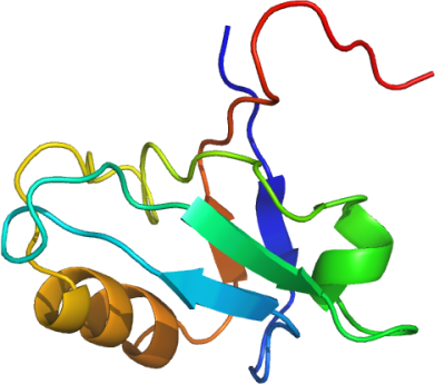
PDB:2V1W
Revision
Revision Type:created
Revised by:created
Revision Date:created
Entry Clone Accession:gi|19923181
Entry Clone Source:MGC
SGC Clone Accession:PDLIM4A-c001
Tag:N-terminal hexahistidine tag followed by TEV cleavage site. C-terminal PDZ binding motif.
Host:BL21(DE3)-R3-pRARE2
Construct
Prelude:
Sequence:smPHSVTLRGPSPWGFRLVGGRDFSAPLTISRVHAGSKAALAALCPGDLIQAINGESTELMTHLEAQNRIKGCHDHLTLSVSRPEGESDL
Vector:pNIC28-Bsa4.
Growth
Medium:LB
Antibiotics:
Procedure:A glycerol stock was used to innoculate a starter culture of 40 ml LB containing 50 µg/ml kanamycin and 34 µg/ml chloramphenicol. The next day 14 mls of the starter culture was used to innoculate 1 litre of TB containing 50 µg/ml kanamycin. The culture was grown at 37°C overnight as a starter culture for a 1 litre growth. The large scale growth was grown at 37°C for approximately 6 hours when the temperature was lowered to 22°C. Protein production was induced with the addition of 0.5 mM IPTG and the cells cultured overnight. The next day cells were harvested by centrifugation at 6000 rpm for 15 minutes and resuspended in 25 mls of Lysis buffer before storage in the -80°C freezer.
Purification
Procedure
Column 1: Low pressure chromatography using Bio-Rad Econo column (2.5 cm x 13cm).
Column 2: Gel Filtration using Superdex 200 column
The Ni-NTA columns were prepared as follows. 4 ml of 50% slurry (regenerated x13) was added to a clean gravity column. The resin was equilibrated with 25 ml Lysis/Binding buffer. The lysates were allowed to drip through the column twice, and the flow through collected .The column was washed with 12.5 ml volumes of Lysis/Binding Buffer and 25 ml (2x12.5ml) of Wash Buffer. The bound protein was eluted with 10 mls Elute Buffer and collected into a 15 ml falcon tube.
The sample was filtered through a 1.2 micron and then a 0.45 micron syringe filter. The columns were washed with 2 column volumes of water at a flow rate of 0.5 ml/min and then equilibrated with 5 column volumes of Binding Buffer at a flow rate of 0.8 ml/min. The sample was then loaded on the gel filtration column in Gel Filtration buffer at 1.0 ml/min. Eluted proteins were collected in 1.8 ml fractions. The fractions containing protein were identified on a Coomasie blue stained gel.
Enzymatic treatment: At this stage the purity of the protein was greater than 95% based on SDS-PAGE analysis. The N-terminal hexahistidine tag was removed by TEV protease treatment. The TEV protease, a hexahistidine-tagged construct, was over-expressed and purified in-house. 400 µl of the TEV protease was added to the pooled gel filtration fractions containing protein PDLIM4 and left at 4°C overnight. The following steps were carried out to remove the cleaved products and TEV protease. Place 200 µl of 50 % Ni-NTA agarose in a 1.5 ml eppendorf tubes, add 1ml of Gel Filtration Buffer mix, spin down and remove buffer. Repeat this resin wash step once. Add the TEV treated protein sample to the resin and mix for 60 mins at 4°C. Finally spin down resin and collect the supernatant which contains the cleaved PDLIM4. The sample was then concentrated to 5.9 mg/ml using a 10 kD cutoff spin concentrator before storage in a -80°C freezer.
Extraction
Procedure
PMSF (final concentration 0.2 mM) in 10 ml of Lysis/ Binding Buffer was added to the thawed cell pellet (approximately 35 mls volume) and mixed by inversion. The cell pellet was lysed by passing it three times through the EmulsiFlex C5 high pressure homogeniser (Evestin), collecting a final volume of approximately 50 mls. PEI was added to a final concentration of 0.15%, mixed by inversion and the cell debris and precipitated DNA were spun down at 21,500rpm for 1hr.
Concentration:
Ligand
MassSpec:After hexahistidine tag removal: Expected 9571.8; Measured 9570.7.
Crystallization:Crystals grew from a 1:2 ratio mix of PDLIM4-to-reservoir ( 20% PEG 10K; 0.20M MgCl2; 0.1M TRIS pH 8.5 ).
NMR Spectroscopy:
Data Collection:1.9Å; X-ray source: Synchrotron SLS- SLS-X10.
Data Processing: