PTK9L
PDB:2VAC
Revision
Revision Type:created
Revised by:created
Revision Date:created
Entry Clone Accession:gi|6005846
Entry Clone Source:MGC
SGC Clone Accession:PTK9LA-c013
Tag:mhhhhhhssgvdlgtenlyfq*s(m) TEV-cleavable (*) N-terminal his6 tag.
Host:BL21(DE3)-R3-pRARE2 (A homemade phage resistant version of BL21(DE3) containing the pRARE2 plasmid from Rosetta II (DE3) cells).
Construct
Prelude:
Sequence:MHHHHHHSSGVDLGTENLYFQ^smGIHATEELKEFFAKARAGSVRLIKVVIEDEQLVLGASQEPVGRWDQDYDRAVLPLLDAQQPCYLLYRLDSQNAQGFEWLFLAWSPDNSPVRLKMLYAATRATVKKEFGGGHIKDELFGTVKDDLSFAGYQKH
Vector:pNIC28-Bsa4.
Growth
Medium:
Antibiotics:
Procedure:Glycerol stock preparation: A number of colonies from the transformation were used to innoculate 1 ml of LB media containing 50 µg/ml kanamycin and 34 µg/ml chloramphenicol, which was placed in a 37°C shaker overnight. The next day glycerol stocks were prepared from this overnight culture.
A glycerol stock was used to innoculate 10 ml of TB media containing 50 µg/ml kanamycin, which was placed in a 37°C shaker overnight. The next day this starter culture was used to innoculate 1L of TB media containing 50 µg/ml kanamycin. After growth at 37°C until the OD600 was greater than 2, the temperature was reduced to 18°C and the cells were induced by the addition of 0.5 mM IPTG. The expression was continued overnight. Two of these 1L growths were combined for the subsequent protein purification. Cells were spun and frozen at -80°C.
Purification
Procedure
Column 1: HisTrap 5ml.
Column 1: 2x HiPrep 26/10 desalting columns in series.
The protein was purified using an AktaExpress system.
Column 1 Procedure: The clarified cell extract was passed through the column at a flow rate of 4 ml/min. The column was then washed Binding Buffer until a stable UV baseline was achieved. The column was then washed with Wash Buffer until a stable UV baseline was achieved. The protein was eluted with 25 ml of Elution Buffer.
TEV protease digestion: The desalted fractions containing PTK9LA were pooled and 700 µl of TEV protease solution (about 1 mg/ml) was added. The digestion was left overnight at 4°C. After 24 hours the TEV protease digestion had not proceeded to completion so an additional 200 µl of TEV protease solution was added and the digestion left for an additional 24 hours.
Rebinding of impurities to Ni-NTA: The protein was mixed with Ni-NTA resin (4 ml, pre-equilibrated into Desalting Buffer) at 4°C for 45 minutes. The resin was spun down and the supernatent filtered through a 0.45 µM syringe filter.
Extraction
Procedure
The cells were thawed and resuspended in 120 ml of Lysis buffer. The resuspended cells were passed 5 times through an Emulsiflex C5 high-pressure homogeniser, collecting a final volume of approximately 170 ml after dilution with Lysis Buffer. PEI was added to a final concentration of 0.2 % and the cell debris and precipitated DNA were spun down (16000 rpm, JLA 16.250 rotor, 38400 x g, 70 min). The supernatent was filtered through a 0.45 µM syringe filter.
Concentration:The TEV protease cleaved PTK9LA was concentrated to 20.8 mg/ml (measured using a nanodrop spectrometer and a calculated extinction coefficient at 280 nm), distributed into aliquots and frozen at -80°C.
Ligand
MassSpec:Measured: 15163.7;
Expected: 15162.1.
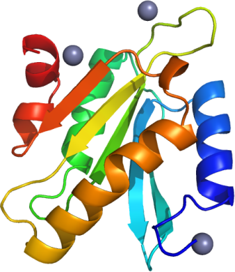
Crystallization:Crystals grew from a 1:1 ratio of protein to precipitant solution (0.01M ZnCl 2 , 0.1M Tris pH 8.0, 20% PEG 6000, 10% Ethylene Glycol), using the vapour diffusion method.
NMR Spectroscopy:
Data Collection:Crystals were cryo-protected by equilibration into precipitant solution containing 25% ethylene glycol, and then flash frozen in liquid nitrogen. Data was collected on a Rigaku FR-E rotating anode X-ray source at 1.7 Å resolution.
Data Processing: