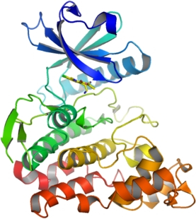
CLK1
PDB:2VAG
Revision
Revision Type:created
Revised by:created
Revision Date:created
Entry Clone Accession:n/a
Entry Clone Source:synthetic
SGC Clone Accession:
Tag:Tag sequence: mhhhhhhssgvdlgtenlyfq*s(m). TEV-cleavable (*) N-terminal hexaHis tag.
Host:BL21 (DE3)
Construct
Prelude:
Sequence:mhhhhhhssgvdlgtenlyfqSMHLICQSGDVLSARYEIVDTLGEGAFGKVVECIDHKAGGRHVAVKIVKNVDRYCEAARSEIQVLEHLNTTDPNSTFRCVQMLEWFEHHGHICIVFELLGLSTYDFIKENGFLPFRLDHIRKMAYQICKSVNFLHSNKLTHTDLKPENILFVQSDYTEAYNPKIKRDERTLINPDIKVVDFGSATYDDEHHSTLVSTRHYRAPEVILALGWSQPCDVWSIGCILIEYYLGFTVFPTHDSKEHLAMMERILGPLPKHMIQKTRKRKYFHHDRLDWDEHSSAGRYVSRACKPLKEFMLSQDVEHERLFDLIQKMLEYDPAKRITLREALKHPFFDLLKKSI
Vector:pLIC- SGC1
Growth
Medium:
Antibiotics:
Procedure:Growth medium, induction protocol: 1 ml from a 10 ml overnight culture in LB, 100 µg/ml ampicillin was used to inoculate 1 litre of LB medium containing 100 µg/ml ampicillin. Cultures were grown at 37°C until they reached an OD600of 0.3 and then cooled to 18°C. Expression was induced for 4 hours using 1 mM IPTG at an OD600 of 0.8. The cells were collected by centrifugation, transferred to 50 ml tubes, resuspended in 30 ml binding buffer, and frozen. Binding buffer: 50 mM HEPES pH 7.5, 500 mM NaCl, 5% Glycerol.
Purification
Procedure
Column 1 : Ion exchange - Nucleic acid removal. DEAE cellulose (DE52, Whatman), 10 g of resin in 2.5 x 20 cm column. The resin was hydrated in 2.5M NaCl, then washed with 20 ml binding buffer prior to loading the sample.Buffers: Binding bufferProcedure: Supernatant was applied at gravity flow, followed by a wash with 50 ml binding buffer. The column flow-through was collected.Column 2: Ni-affinity. Ni-NTA (Qiagen), 5 ml of 50% slurry in 1.5 x 10 cm column, washed with binding buffer.Buffers : Binding buffer: 50 mM HEPES pH 7.5, 500 mM NaCl, 5% Glycerol.Wash buffer: 50 mM HEPES pH 7.5, 500 mM NaCl, 20 mM Imidazole, 5% glycerolElution buffer: 50 mM HEPES pH 7.5, 500 mM NaCl, 50 to 250 mM Imidazole , 5% GlycerolProcedure: The flowthrough from column 1 was loaded by gravity flow on the Ni-NTA column. The column was then washed with 3 x 10 ml wash buffer at gravity flow. The protein was eluted by gravity flow by applying 5-ml portions of elution buffer with increasing concentration of imidazole (50 mM, 100 mM, 150 and 250 mM); fractions were collected until essentially all protein was eluted. After elution DTT was added to a final concentration of 10 mM.Enzymatic treatment : (Dephosphorylation and His tag cleavage) Samples containing CLK1 were pooled and 20 µg GST-lambda phosphatase and 20 µg TEV protease added for overnight incubation at 4°C: protein solution contained 10 mM DTT and 0.05 mM MnCl2Column 3: Size Exclusion ChromatographyBuffers: Fractions containing CLK1 collected from IMAC were concentrated and directly applied to a S75 16/60 HiLoad gel filtration column equilibrated in 50 mM Hepes pH 7.5, 500 mM NaCl, 50 mM L-glutamic acid, 50 mM L-arginineProcedure : AKTA-prime
Extraction
Procedure
Extraction buffer, extraction method : The frozen cells were thawed on ice and binding buffer (plus 1 mM PMSF) added to a final volume of 50 ml. Cells were lysed using a high pressure cell disruptor. The lysate was centrifuged at 17,000 RPM for 30 minutes and the supernatant collected for purification.
Concentration:Protein concentration: Conditions for the concentration of the protein were found using a thermofluor analysis of the stability in HEPES (pH 7.5) buffer with different salts/additives. Following a report by A.P. Golovanov et al. (2004, J. Am. Chem. Soc. 126: 8933-8939), a combination of L-arginine and L-glutamic acid at 50 mM and a high concentration of NaCl (500 mM) was tested among other additives. The thermal stability was measured by monitoring the fluorescence increase of the fluorescent dye SYPRO orange (Molecular Probes). The midpoint of the unfolding transition, Tm, was shifted by the addition of L-arginine/L-glutamic acid by more than 10°C . Small-scale concentrations (VivaSpin PES membrane, 10-kDa cut-off) showed a more than 10-fold increased solubility under these conditions. For setup of crystallization experiments the protein was concentrated from 0.1 mg/ml to 8 mg/ml in the presence of a 4-fold excess of inhibitor and an 8-fold excess of a substrate peptide (AFRREWSPGKEAKK). Protein was buffered in 25 mM HEPES pH 7.5, 250 mM NaCl, 1.5% glycerol, 50 mM L-arginine, 50 mM L-glutamate, 10 mM DTT.
Ligand
ethyl 3-[(E)-2-amino-1-cyanoethenyl]-6,7- dichloro-1-methyl-1H-indole-2-carboxylateMassSpec:Mass spec characterization: LC- ESI -MS TOF confirmed the correct mass expected for this construct.Intact Mass: Masses of purified proteins were confirmed by LC-MS, using an Agilent LC/MSD TOF system with reversed-phase HPLC coupled to electrospray ionisation and an orthogonal time-of-flight mass analyser. Proteins were desalted prior to mass spectrometry by rapid elution off a C3 column with a gradient of 5-95% acetonitrile in water with 0.1% formic acid.
Crystallization:Crystallization: Crystals were grown at 4°C in 150nl sitting drops mixing 100 nl of CLK1 (8 mg/ml) with 50 nl of a solution containing 2.1 M sodium malate pH 7.0.
NMR Spectroscopy:
Data Collection:Data Collection: Resolution: 1.8Å, X-ray source: Synchrotron SLS-X10, single wavelength.
Data Processing: