ACADS
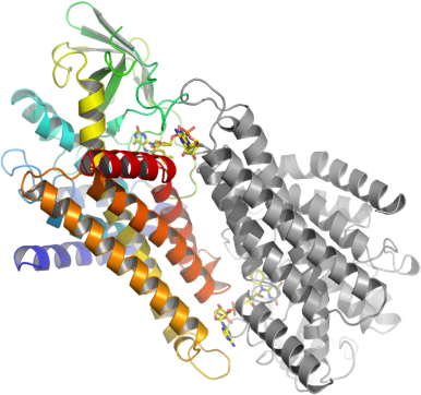
PDB:2VIG
Revision
Revision Type:created
Revised by:created
Revision Date:created
Entry Clone Accession:gi|4557233
Entry Clone Source:MGC
SGC Clone Accession:IMAGE:4842286
Tag:C-terminal, TEV-cleavable(*) Histidine tag + Flag epitope: aenlyfq*shhhhhhdykddddk
Host:BL21(DE3)-R3-pRARE2
Construct
Prelude:
Sequence: Expressed:mQSVELPETHQMLLQTCRDFAEKELFPIAAQVDKEHLFPAAQVKKMGGLGLLAMDVPEELGGAGLDYLAYAIAMEEISRGCASTGVIMSVNNSLYLGPILKFGSKEQKQAWVTPFTSGDKIGCFALSEPGNGSDAGAASTTARAEGDSWVLNGTKAWITNAWEASAAVVFASTDRALQNKSISAFLVPMPTPGLTLGKKEDKLGIRGSSTANLIFEDCRIPKDSILGEPGMGFKIAMQTLDMGRIGIASQALGIAQTALDCAVNYAENRMAFGAPLTKLQVIQFKLADMALALESARLLTWRAAMLKDNKKPFIKEAAMAKLAASEAATAISHQAIQILGGMGYVTEMPAERHYRDARITEIYEGTSEIQRLVIAGHLLRSYRSaenlyfq*shhhhhhdykddddkAfter tag cleavage:mQSVELPETHQMLLQTCRDFAEKELFPIAAQVDKEHLFPAAQVKKMGGLGLLAMDVPEELGGAGLDYLAYAIAMEEISRGCASTGVIMSVNNSLYLGPILKFGSKEQKQAWVTPFTSGDKIGCFALSEPGNGSDAGAASTTARAEGDSWVLNGTKAWITNAWEASAAVVFASTDRALQNKSISAFLVPMPTPGLTLGKKEDKLGIRGSSTANLIFEDCRIPKDSILGEPGMGFKIAMQTLDMGRIGIASQALGIAQTALDCAVNYAENRMAFGAPLTKLQVIQFKLADMALALESARLLTWRAAMLKDNKKPFIKEAAMAKLAASEAATAISHQAIQILGGMGYVTEMPAERHYRDARITEIYEGTSEIQRLVIAGHLLRSYRSaenlyfqSequences in lowercase are derived from the vector; the asterisk indicates the site of cleavage by the TEV protease.
Vector: pNIC-CTHF
Growth
Medium:
Antibiotics:
Procedure:The expression plasmid was transformed into the host strain and plated on LB-agar containing 50 µg/ml kanamycin and 35 µg/ml chloramphenicol. Several colonies were combined to inoculate a 1-ml culture in TB (+ 50 µg/ml kanamycin, 35 µg/ml chloramphenicol). The culture was grown overnight, glycerol was added to 15% v/v (from a 60% stock), and the resulting glycerol stock was frozen at -80°C in 100 µl aliquots. A loopful of cells from the glycerol stock was inoculated into 2x 10-ml of TB medium containing 50 µg/ml kanamycin and 35 µg/ml chloramphenicol and grown overnight at 37°C. 3-L of TB medium (+ 50 mg/ml kanamycin) in UltraYield baffled flasks was inoculated with the overnight cultures. The culture was grown at 37°C until OD600 of 3.2 and then shifted to 18°C. After 30 minutes, IPTG was added to 0.1 mM, and growth continued overnight. The cells were collected by centrifugation then the pellets were scraped out and transferred to 50-ml Falcon tubes and frozen at -80°C.
Purification
Procedure
Column 1: Ni-affinity, HisTrap Crude FF, 5 ml (GE Healthcare)The cell extract was loaded on the column at 4 ml/minute on an AKTA-express system (GE Healthcare). The column was washed with 10 volumes of lysis buffer, 10 volumes of wash buffer, and then eluted with elution buffer at 4 ml/min. The eluted peak of A280 was automatically collected. The protein fractions were yellow throughout purification and crystallization.Column 2: Gel filtration, Hiload 16/60 Superdex S75 prep grade, 120 ml (GE Healthcare)The eluted fraction from the Ni-affinity Histrap column was loaded on the gel filtration column in GF buffer at 0.80 ml/min. Eluted proteins were collected in 2-ml fractions and analyzed on SDS-PAGE.Tag removal: The pooled protein fractions from the gel filtration columns were treated with TEV protease (1/20 w/w) overnight at 4°C. The solution was then passed over a NiNTA column to remove contaminating proteins.
Extraction
Procedure
Frozen cell pellets (47g) were thawed briefly in a bath of warm water (20 - 37°C) then transferred to ice. One volume (i.e. 1 ml for every gram of cells) of 2x lysis buffer was added, followed by 1x lysis buffer to a total volume of 150 ml. The cells were resuspended by agitating and disrupted by high pressure homogenization (20 kpsi). Nucleic acids and cell debris were removed by adding 0.15% PEI (polyethyleneimine) from a 5% (w/v, pH 7.5) stock, stirring for 15 minutes, then centrifugation for 20 minutes at 25,000 x g. The supernatant was then further clarified by filtration (Acrodisc filters, 0.2 mm).
Concentration:The protein was supplemented with 5 mM acetyl-CoA, concentrated in Amicon (30 K) to 26.6 mg/ml and stored at 4°C. The protein concentration was determined spectrophotometrically using ε280=72770.
Ligand
MassSpec:Observed mass 42124 Da, which is 4 Da higher than the predicted 42120.54 Da.
Crystallization:Crystals were grown by vapour diffusion from nanolitre sitting drops. The protein was mixed with a reservoir solution containing 0.20M NH4Cl, 0.1M Tris-HCl pH 8.0, 20.0% PEG 6K, 10.0% EtGly (50 nl protein:100 nl solution), and allowed to equilibrate by vapour diffusion.
NMR Spectroscopy:
Data Collection:Data Collection: Resolution: 2.65Å; X-ray source: Swiss Light source (SLS), beamline X-10.
Data Processing: