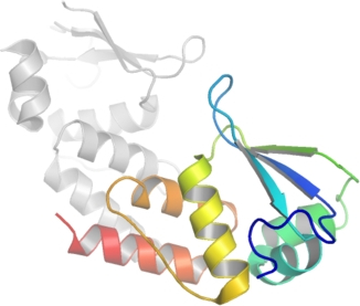
BTBD6
PDB:2VKP
Revision
Revision Type:created
Revised by:created
Revision Date:created
Entry Clone Accession:IMAGE:4824525
Entry Clone Source:MGC
SGC Clone Accession:
Tag: mhhhhhhssgvdlgtenlyfq*s(m) TEV-cleavable (*) N-terminal his6 tag.
Host:BL21(DE3)-R3-pRARE2
Construct
Prelude:
Sequence: mhhhhhhssgvdlgtenlyfq*sMFNNELMADVHFVVGPPGATRTVPAHKYVLAVGSSVFYAMFYGDLAEVKSEIHIPDVEPAAFLILLKYMYSDEIDLEADTVLATLYAAKKYIVPALAKACVNFLETSL
Vector:pNIC28-Bsa4
Growth
Medium:
Antibiotics:
Procedure:The expression plasmid was transformed into the host strain and plated on LB-agar containing 50 µg/ml kanamycin and 35 µg/ml chloramphenicol. Several colonies were combined to inoculate a 1-ml culture in TB (+ 50 µg/ml kanamycin, 35 µg/ml chloramphenicol). The culture was grown overnight, glycerol was added to 15% v/v (from a 60% stock), and the resulting glycerol stock was frozen at -80°C in 100 µl aliquots. A loopful of cells from the glycerol stock was inoculated into 6x 10-ml of LB medium containing 100 µg/ml kanamycin and 35 µg/ml chloramphenicol and grown overnight at 37°C. Cultures were harvested by centrifugation and washed twice with M9 minimal medium and resuspended in 10 ml M9 minimal medium. 6x 1L M9 minimal medium (containing 0.4% glucose, 2mM MgSO4, 0.1mM CaCl2, 50 µg/ml kanamycin) were each inoculated with 10 ml resuspended cells and grown in 2.5L UltraYield baffled flasks until OD600 of 0.80. Selenomethioine was added to 25mg/L along with leucine, isoleucine and valine to 50mg/L and lysine, threonine, and phenylalanine to 100mg/L (all amino acids dissolved in 0.2M HEPES pH 7.5). Cultures were grown for a further 1.5 hours until OD600 of 1.2 and then cooled to 18°C for 1 hour. Additional selenomethioine was added (final total concentration of 75mg/L) and IPTG was added to 0.1 mM, and growth continued at 18°C overnight. The cells were collected by centrifugation then the pellets were scraped out and transferred to 50-ml Falcon tubes and frozen at -80°C.
Purification
Procedure
Column 1 : Ni-affinity, HisTrap Crude FF, 5 ml (GE Healthcare)
Solutions: Affinity buffer: 50 mM HEPES buffer, pH 7.5, 500 mM NaCl, 10 mM imidazole, 5% Glycerol, 0.5 mM TCEP. Wash buffer: 50 mM HEPES buffer, pH 7.5, 500 mM NaCl, 30 mM imidazole, 5% Glycerol, 0.5 mM TCEP. Elution buffer: 50 mM HEPES buffer, pH 7.5, 500 mM NaCl, 250 mM imidazole, 5% Glycerol, 0.5 mM TCEP.
Procedure: The cell extract was loaded on the column at 0.8 ml/minute on an AKTA-express system (GE Healthcare). The column was washed with 10 volumes of lysis buffer, 10 volumes of wash buffer, and then eluted with elution buffer at 0.8 ml/min. The eluted peak of A280 was automatically collected.
Column 2 : Gel filtration, Hiload 16/60 Superdex S75 prep grade, 120 ml (GE Healthcare)
GF buffer: 10 mM HEPES, pH 7.5, 500 mM NaCl, 5% Glycerol, 0.5 mM TCEP.
Procedure: The eluted fractions from the Ni-affinity Histrap column was loaded on the gel filtration column in GF buffer at 1.2 ml/min. Eluted proteins were collected in 2-ml fractions and analyzed on SDS-PAGE.
Enzymatic treatment and purification: The N-terminal His6-tag was cleaved by incubating the protein overnight with TEV protease (rotating at 4°C). Cleaved protein was purified by batch binding on 2ml pre-equilibriated 50% Ni-NTA bead solution (rotating at 4°C) for 1 hour then the column poured. Elution was done in GF buffer and eluate collected.
Extraction
Procedure
Frozen cell pellets (28g) were thawed briefly in a bath of warm water (20 ~ 37°C) then transferred to ice. One volume (i.e. 1 ml for every gram of cells) of 2x lysis buffer was added, followed by 1x lysis buffer to a total volume of 50-ml. The cells were resuspended by agitating and disrupted by high pressure homogenization (20 kpsi). Nucleic acids and cell debris were removed by adding 0.15% PEI (polyethyleneimine) from a 5% (w/v, pH 7.5) stock, stirring for 15 minutes, then centrifugation for 20 minutes at 25,000 x g . The supernatant was then further clarified by filtration (Acrodisc filters, 0.2 µm).
Concentration:The cleaved purified protein was concentrated in a VivaSpin4 (5 K MWCO) to 9.7 mg/ml and stored at 4°C. The protein concentration was determined spectrophotometrically using ε280 = 10430.
Ligand
MassSpec:
Crystallization:Crystals were obtained using the vapor diffusion method. The selenomethionine-labelled protein was concentrated in gel fitration buffer to a protein concentration of 9.7 mg/ml. Sitting drops comprising 135 nl of the concentrated protein mixed with 165nl of a well solution (0.8M potassium citrate, 0.1M sodium cacodylate pH 6.5) were equilibrated against well solution at 4°C. Hexagonal rods appeared within 2-3 days.
NMR Spectroscopy:
Data Collection:Crystals were cryo-protected using the well solution supplemented with an additional 30% ethylene glycol and flash frozen in liquid nitrogen. Diffraction data were collected from a single crystal at the Se-peak (λ=0.97874Å) and at a low-energy remote (native, λ=0.98248Å) on beamline X10SA at the SLS (Swiss light source). The structure was solved using SAD and refined to 1.9Å.
Data Processing: