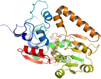
RTN4IP1 + NADPH
PDB:2VN8
Revision
Revision Type:created
Revised by:created
Revision Date:created
Entry Clone Accession:IMAGE:4309263
Entry Clone Source:MGC
SGC Clone Accession:Tag:N-terminal TEV-cleavable (at *) his-tag with the following sequence: mhhhhhhssgvdlgtenlyfq*s
Host:BL21(DE3)-R3-pRARE2
Construct
Prelude:Sequence:mhhhhhhssgvdlgtenlyfq*SMAWVIDKYGKNEVLRFTQNMMMPIIHYPNEVIVKVHAASVNPIDVNMRSGYGATALNMKRDPLHVKIKGEEFPLTLGRDVSGVVMECGLDVKYFKPGDEVWAAVPPWKQGTLSEFVVVSGNEVSHKPKSLTHTQAASLPYVALTAWSAINKVGGLNDKNCTGKRVLILGASGGVGTFAIQVMKAWDAHVTAVCSQDASELVRKLGADDVIDYKSGSVEEQLKSLKPFDFILDNVGGSTETWAPDFLKKWSGATYVTLVTPFLLNMDRLGIADGMLQTGVTVGSKALKHFWKGVHYRWAFFMASGPCLDDIAELVDAGKIRPVIEQTFPFSKVPEAFLKVERGHARGKTVINVV
Vector:pNIC28-Bsa4
Growth
Medium:Antibiotics:Procedure:TB. 10ml of overnight culture was added into 1L TB with 50 µg/ml of Kanamycin and 34 mg/ml of Chloramphenicol (total 12L). The cells were cultured at 37°C until the OD reached 1.360 and then decreased the temperature to 18°C. IPTG was added at 0.5mM (final concentration) and kept the culture at 18°C for overnight.
For Selenomethionine labelling, the plasmid was transformed into B834 (DE3) cells. Single colony was cultured in 1000 ml of LB media with 50 µg/ml of Kanamycin at 30°C overnight. The cells then were washed and cultured in 12L MD media with 40mg of Selenomethionine/L at 37°C. When the OD reached 1.0, 0.5 mM (final concentration) of IPTG was added and the temperature was decreased to 25°C for overnight culture.
Purification
ProcedureColumn 1: Ni-NTA
Buffers: Binding buffer: 500 mM NaCl, 5% Glycerol, 50 mM HEPES pH 7.5, 40 mM Imidazole; Washing Buffer: 500 mM NaCl, 5% Glycerol, 50 mM HEPES pH 7.5, 40 mM Imidazole; Elution Buffer I: 500 mM NaCl, 5% Glycerol, 50 mM HEPES pH 7.5, 60 mM Imidazole; Elution Buffer II: 500 mM NaCl, 5% Glycerol, 50 mM HEPES pH 7.5, 80 mM Imidazole; Elution Buffer III: 500 mM NaCl, 5% Glycerol, 50 mM HEPES pH 7.5, 125 mM Imidazole; Elution Buffer VI: 500 mM NaCl, 5% Glycerol, 50 mM HEPES pH 7.5, 250 mM Imidazole.
Procedure: The column was packed by 4 ml of Ni-NTA slurry and equilibrated with 15 ml of binding buffer. The supernatant was loaded onto the column and the flow-through was collected. The column was washed with 2x20 ml of washing buffer. The protein was eluted with 5 ml of elution buffer I, II & III respectively and then 8 ml of elution buffer VI.
Column 2: Superdex 200 Hiload 16 60
Buffers: 500 mM NaCl, 5% Glycerol, 50 mM HEPES pH 7.5, 0.5 mM TCEP.
Procedure: AKTA Purifier was used and run at 4°C. Fractions were analyzed by SDS -PAGE and the most purified fractions were collected.
Column 3: Hi-Trap 5ml SP-HP column.
Buffers: Low salt buffer: 20 mM Hepes, pH 7.5, 100 mM NaCl; High salt buffer: 20 mM Hepes, pH 7.5, 2 M NaCl.
Procedure: The protein was applied to 5ml SP-HP column in low salt buffer and eluted from the column by a linear gradient with high salt buffer.
Extraction
Procedure500 mM NaCl, 5% Glycerol, 50 mM HEPES pH 7.5, 5 mM Imidazole. Complete Protease Inhibitor Cocktail Tablets (Roche) were added (one tablet/50ml buffer). The cells were harvested by centrifugation at 4,000 g for 10 min. The pellet from 1 L culture was resuspended in 25 ml of extraction buffer. The sample was homogenized by using the EmulsiFlex-05 homogenizer (Glen Creston) and then centrifuged at 37505 g. The supernatant was kept for further purification.
Concentration:8.4 mg/ml
LigandNADPH
MassSpec:41220 (expected); 41783 (Selenomethionine labelled, experimental mass as expected for fully labelled protein)
Crystallization:All crystallizations were carried out using sitting drop vapour diffusion at 4°C. Native crystals were grown by mixing protein solution (8mg/ml) with 0.85-1.0x dilution of reservoir solution at a ratio of 120nl protein:180nl reservoir and equilibrating against the same reservoir solution. The reservoir solution comprised 40% MPD, 0.15M ammonium acetate, 0.1M sodium citrate pH5.6. Crystallization of SeMet-substituted protein was performed by matrix-seeding with crushed native crystals into 200nl drops composed of equal volumes of SeMet protein (8mg/ml) and reservoir solution (40% MPD, 0.25M ammonium acetate, 0.1M sodium citrate pH5). In both cases, 5mM NADPH was added to the protein prior to crystallization.
NMR Spectroscopy:Data Collection:Native and Se-SAD data were collected at beamline X10SA at the Swiss Light Source (SLS). Initial phases were determined using SAD data collected from crystals of selenomethionine-substituted protein at the Se peak (λ >= 0.979 Å). Phase extension, two-fold NCS averaging and solvent flattening in DM allowed the entire molecule to be traced. The structure has been refined to 2.1 Å resolution.
Data Processing: