MYNN
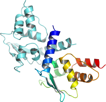
PDB:2VPK
Revision
Revision Type:created
Revised by:created
Revision Date:created
Entry Clone Accession:gi|8923885
Entry Clone Source:MGC
SGC Clone Accession:IMAGE:5201998
Tag:Tag sequence: mhhhhhhssgvdlgtenlyfq*s(m) TEV-cleavable (*) N-terminal his6 tag.
Host:BL21(DE3)-R3-pRARE2 (previously known as Rosetta)
Construct
Prelude:
Sequence:mhhhhhhssgvdlgtenlyfq*smSHHCEHLLERLNKQREAGFLCDCTIVIGEFQFKAHRNVLASFSEYFGAIYRSTSENNVFLDQSQVKADGFQKLLEFIYTGTLNLDSWNVKEIHQAADYLKVEEVVTKCKIKMED
Vector:pNIC28-Bsa4
Growth
Medium:
Antibiotics:
Procedure:The expression plasmid was transformed into the host strain and plated on LB-agar containing 50 µg/ml kanamycin and 35 µg/ml chloramphenicol. Several colonies were combined to inoculate a 1ml culture in TB (+ 50 µg/ml kanamycin, 35 µg/ml chloramphenicol). The culture was grown overnight, glycerol was added to 15% v/v (from a 60% stock), and the resulting glycerol stock was frozen at -80°C in 100 µl aliquots. A loopful of cells from the glycerol stock was inoculated into 6x 10-ml of TB medium containing 100 µg/ml kanamycin and 35 µg/ml chloramphenicol and grown overnight at 37°C. Cultures were grown for a further 1.5 hours until OD600 of 3.6 and then cooled to 18°C for 1 hour. IPTG was added to 0.1 mM, and growth continued at 18°C overnight. The cells were collected by centrifugation then the pellets were scraped out and transferred to 50-ml Falcon tubes and frozen at -80°C.
Purification
Procedure
Column 1 : Ni-affinity, HisTrap Crude FF, 5 ml (GE Healthcare)Buffers: Affinity buffer: 50 mM HEPES buffer, pH 7.5, 500 mM NaCl, 10 mM imidazole, 5% Glycerol, 0.5 mM TCEP. Wash buffer: 50 mM HEPES buffer, pH 7.5, 500 mM NaCl, 30 mM imidazole, 5% Glycerol, 0.5 mM TCEP. Elution buffer: 50 mM HEPES buffer, pH 7.5, 500 mM NaCl, 250 mM imidazole, 5% Glycerol, 0.5 mM TCEP.Procedure: The cell extract was loaded on the column at 4 ml/minute on an AKTA-express system (GE Healthcare). The column was washed with 10 volumes of lysis buffer, 10 volumes of wash buffer, and then eluted with elution buffer at 4 ml/min. The eluted peak of A280 was automatically collected.Column 2 : Gel filtration, Hiload 16/60 Superdex S75 prep grade, 120 ml (GE Healthcare)GF buffer: 10 mM HEPES, pH 7.5, 500 mM NaCl, 5% Glycerol, 0.5 mM TCEP.Procedure: The eluted fractions from the Ni-affinity Histrap column was loaded on the gel filtration column in GF buffer at 1.2 ml/min. Eluted proteins were collected in 2-ml fractions and analyzed on SDS-PAGE.Enzymatic treatment and purification: The N-terminal His6-tag was cleaved by incubating the protein overnight with TEV protease (rotating at 4°C). Uncleaved protein and other impurities were removed by batch binding on a 1 ml pre-equilibriated 50% Ni-NTA bead solution (rotating at 4°C) for 1 hour. The resin was poured into a column and the cleaved protein was eluted with GF buffer.
Extraction
Procedure
Lysis buffer: 50 mM HEPES buffer, pH 7.5, 500 mM NaCl, 10 mM imidazole, 5% glycerol, 0.5 mM TCEP, 1x Protease Inhibitors Cocktail Set VII (Calbiochem, 1/1000 dilution), and 15 units/ml Benzonase. 2x Lysis buffer contains the same components at double concentration.Procedure: Frozen cell pellets (47.5g) were thawed briefly in a bath of warm water (20 - 37°C) then transferred to ice. One volume (i.e. 1 ml for every gram of cells) of 2x lysis buffer was added, followed by 1x lysis buffer to a total volume of 100 ml. The cells were resuspended by agitating and disrupted by high pressure homogenization (20 kpsi). Nucleic acids and cell debris were removed by adding 0.15% PEI (polyethyleneimine) from a 5% (w/v, pH 7.5) stock, stirring for 15 minutes, then centrifugation for 20 minutes at 25,000 x g. The supernatant was then further clarified by filtration (Acrodisc filters, 0.2 µm).
Concentration:The cleaved purified protein was concentrated in a VivaSpin500 (5 K MWCO) to 6.2 mg/ml and stored at 4°C. The protein concentration was determined spectrophotometrically.
Ligand
MassSpec:Observed mass without histidine tag, 13439.7 (calculated mass without histidine tag, 13439).
Crystallization:Crystals were obtained using the vapor diffusion method. The protein was concentrated in gel filtration buffer to a protein concentration of 6.2 mg/ml. Sitting drops comprising 50 nl of the concentrated protein mixed with 100nl of a well solution (0.20M Na/KPO4; 0.1M BTProp pH 6.5; 20.0% PEG 3350; 10.0% Ethylene Glycol) were equilibrated against well solution at 4°C. Crystals appeared within 2-3 days.
NMR Spectroscopy:
Data Collection:Crystals were cryo-protected using the well solution supplemented with an additional 25% ethylene glycol and flash frozen in liquid nitrogen. Diffraction data were collected from a single crystal on beamline X10SA at the SLS (Swiss light source). The structure was solved by molecular replacement and refined to 2.0 Å.
Data Processing: