DHP
PDB:2VR2
Revision
Revision Type:created
Revised by:created
Revision Date:created
Entry Clone Accession:BC034395
Entry Clone Source:Mammalian Gene Collection
SGC Clone Accession:DHPA-k010
Tag:N-terminal hexahistidine tag with integrated TEV protease cleavage site: mhhhhhhssgvdlgtenlyfq*sm
Host:E.coli BL21(DE3) (Novagen)
Construct
Prelude:Sequence:mhhhhhhssgvdlgtenlyfq*smAAPSRLLIRGGRVVNDDFSEVADVLVEDGVVRALGHDLLPPGGAPAGLRVLDAAGKLVLPGGIDTHTHMQFPFMGSRSIDDFHQGTKAALSGGTTMIIDFAIPQKGGSLIEAFETWRSWADPKVCCDYSLHVAVTWWSDQVKEEMKILVQDKGVNSFKMFMAYKDLYMVTDLELYEAFSRCKEIGAIAQVHAENGDLIAEGAKKMLALGITGPEGHELCRPEAVEAEATLRAITIASAVNCPLYIVHVMSKSAAKVIADARRDGKVVYGEPIAASLGTDGTHYWNKEWHHAAHHVMGPPLRPDPSTPDFLMNLLANDDLTTTGTDNCTFNTCQKALGKDDFTKIPNGVNGVEDRMSVIWEKGVHSGKMDENRFVAVTSTNAAKIFNLYPRKGRIAVGSDADIVIWDPKGTRTISAKTHHQAVNFNIFEGMVCHGVPLVTISRGKVVYEAGVFSVTAGDGKFIPRKPFAEYIYKRIKQRDRTCTPTPVERAPYKGEVATLKSRVTKEDATAGTRKQAHP
Vector:pNIC-Bsa4
Growth
Medium:Antibiotics:Procedure:Cells from a glycerol stock were grown in 100 ml LB supplemented with, 50 µg/ml kanamycin at 30 ºC overnight. 20 ml of the overnight culture was used to inoculate 1.5 l TB supplemented with 8 g/l glycerol, 50 µg/ml kanamycin and approximately 100 µl BREOX FMT 30 anti-foam solution (Cognis Performance Chemicals UK Ltd). The culture was grown in a LEX bioreactor system (Harbinger Biotechnology) at 37 ºC until OD600 reached ~1. The bottle was down-tempered to 18 ºC over a period of 1 hour before target expression was induced by addition of 0.5 mM IPTG. Expression was allowed to continue overnight and cells were harvested the following morning by centrifugation (4,700 x
g, 20 min, 4 ºC). The resulting cell pellet (24.8 g wet cell weight) was stored at -80 ºC.
Purification
ProcedureColumns
IMAC: Ni-charged 1 ml HiTrap Chelating HP (GE Healthcare)
Gel filtration column: HiLoad 16/60 Superdex 200 Prep Grade (GE Healthcare)
Procedure
Purification of the protein was performed as a two step process on an ÄKTAxpress system (GE Healthcare). Prior to purification, columns were equilibrated with IMAC wash1 buffer and gel filtration buffer, respectively. The filtered lysate was loaded onto two HiTrap Chelating columns connected in series and washed with IMAC wash1 buffer followed by IMAC wash2 buffer. Bound protein was eluted from the IMAC columns with IMAC elution buffer and automatically loaded onto the gel filtration column. Fractions containing the target protein were pooled. The protein was subsequently concentrated using an Amicon Ultra-15 centrifugal filter device with 30,000 NMWL (Millipore) to 18.2 mg/ml in a volume of 1.0 ml. The identity of the protein was confirmed by mass spectrometry.
Extraction
ProcedureThe cell pellet was quickly thawed and resuspended in lysis buffer (1.5 ml/g cell pellet) supplemented with one tablet of Complete EDTA-free protease inhibitor (Roche Applied Science), 1000 U Benzonase (Merck) and a knife edge of lysozyme (Sigma). Cells were disrupted by high-pressure homogenization (TC5-0612W-332, Stansted fluid power LTD) and cell debris was removed by centrifugation (49,000 x
g, 30 min, 4 ºC).
Concentration:LigandMassSpec:Crystallization:Crystals were obtained by the sitting drop vapour diffusion method in a 96-well plate. Prior to setting up the plate, the protein solution was mixed with chymotrypsin (Sigma-Aldrich) in a protease:protein ratio of 1:100 (w/w). 0.2 µl of the protein solution (18.2 mg/ml) was mixed with 0.1 µl of well solution consisting of 0.1 M HEPES pH 7.5 and 70% MPD. The plate was incubated at 4 ºC and crystals appeared between 7 and 14 days. The crystal was flash-frozen in liquid nitrogen without any cryo solution added. Overnight cleavage of the protein with chymotrypsin followed by MS analysis showed that the most probable sequence in the crystal was:
AAPSRLLIRGGRVVNDDFSEVADVLVEDGVVRALGHDLLPPGGAPAGLRV
LDAAGKLVLPGGIDTHTHMQFPFMGSRSIDDFHQGTKAALSGGTTMIIDFAI
PQKGGSLIEAFETWRSWADPKVCCDYSLHVAVTWWSDQVKEEMKILVQD
KGVNSFKMFMAYKDLYMVTDLELYEAFSRCKEIGAIAQVHAENGDLIAEGAK
KMLALGITGPEGHELCRPEAVEAEATLRAITIASAVNCPLYIVHVMSKSAAKVI
ADARRDGKVVYGEPIAASLGTDGTHYWNKEWHHAAHHVMGPPLRPDPST
PDFLMNLLANDDLTTTGTDNCTFNTCQKALGKDDFTKIPNGVNGVEDRM
SVIWEKGVHSGKMDENRFVAVTSTNAAKIFNLYPRKGRIAVGSDADIVIWDP
KGTRTISAKTHHQAVNFNIFEGMVCHGVPLVTISRGKVVYEAGVFSVTAGDG
KFIPRKPFAEYIYKRIKQRDRTCTPTPVERAPYKGEVATL
with a molecular weight of 54435.2 Da.
NMR Spectroscopy:Data Collection:Data was collected at ESRF (ID14-2).
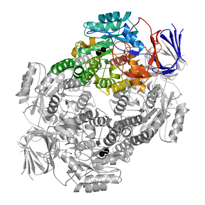
Data Processing:The crystal belonged to space group P6422 with the cell parameters 89.65 89.65 219.1 90 90 120. Data was processed and scaled using XDS and XSCALE. The structure was solved by molecular replacement using MOLREP with the structure of dihydropyrimidinase from slime mold (PDB-code: 2FTW) as search model. Simulated annealing was run in CNS, manual model building was done with COOT and the structure was refined with REFMAC5 using one TLS group covering all protein atoms in the end of the refinement. Data in the interval 20 Â 2.8 Å resolution was used and final values for R and Rfree were 20.3% and 25.1% respectively. The coordinates for the crystal structure were deposited in the Protein Data Bank, accession code 2VR2.