TBCTPS
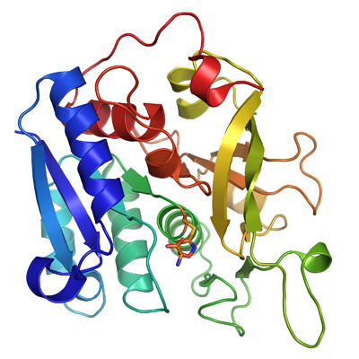
PDB:2W7T
Revision
Revision Type:created
Revised by:created
Revision Date:created
Entry Clone Accession:Similar to GI 115504067 but with one amino acid substitution (K282E)
Entry Clone Source:Through collaboration
SGC Clone Accession:Tag:N-terminal hexahistidine tag with integrated TEV protease cleavage site: mhhhhhhssgvdlgtenlyfq*sm
Host:E.coli BL21(DE3) R3 pRARE, where R3 denotes a derivative of BL21(DE3) resistant to a strain of T1 bacteriophage (SGC Oxford) and the pRARE plasmid originating from the Rosetta strain (Novagen) supplies tRNAs for rare codons.
Construct
Prelude:Sequence:mhhhhhhssgvdlgtenlyfq*smYMSNPTVRIAFVGKYLQDAGDTYFSVLQCFEHCQIALQVRLDILYVDSEELEGPNADEARKALLGCDGIFVPGGFGNRGVDGKCAAAQVARMNNIPYFGVCLGMQVAVIELSRNVVGWSDANSEEFNKESTHQVVRIMDCDRNKMGANMHLGACDVYIVEKSSIMAKIYSKSNIVVERHRHRYEVNTAYFEDLRKAGLCISAVTDPTFSSRCRVEAVENPSLRFFLAVQFHPEFISTPMDPAPTYLSFMAAAAKKDYVWPQKCSQRRLKQA
Vector:pNIC-Bsa4
Growth
Medium:Antibiotics:Procedure:Cells from a glycerol stock were grown in 40 ml TB supplemented with 8 g/l glycerol, 50 µg/ml kanamycin and 34 µg/ml chloramphenicol at 30 ºC overnight. The overnight culture was used to inoculate 4 x 750 ml TB supplemented with 8 g/l glycerol, 50 µg/ml kanamycin, 34 µg/ml chloramphenicol and approximately 500 µl 204 Antifoam A6426 (Sigma) per 750 ml culture. The cultures were grown in TunAir flasks at 37 ºC until OD600 reached ~1.4. The flasks were down-tempered to 15 ºC over a period of 1 hour before target expression was induced by addition of 0.5 mM IPTG. Expression was allowed to continue over the weekend and cells were harvested by centrifugation (5,500 x
g, 10 min, 4 ºC). The resulting cell pellet (88 g wet cell weight) was resuspended in lysis buffer (1.4 ml/g cell pellet), supplemented with 4000 U Benzonase (Merck) and 2 tablets of Complete EDTA-free protease inhibitor (Roche Applied Science). The cell suspension was stored at -80 ºC.
Purification
BuffersIMAC wash1 buffer: 20 mM HEPES, 500 mM NaCl, 10% glycerol, 10 mM imidazole, 0.5 mM TCEP, pH 7.5
IMAC elution buffer: 20 mM HEPES, 500 mM NaCl, 10% glycerol, 500 mM imidazole, 0.5 mM TCEP, pH 7.5
Gel filtration (GF) buffer: 20 mM HEPES, 300 mM NaCl, 10% glycerol, 0.5 mM TCEP, pH 7.5
ProcedureColumnsIMAC 1: Ni-charged 5 ml HiTrap Chelating HP (GE Healthcare)
IMAC 2: Ni-charged 1 ml HiTrap Chelating HP (GE Healthcare)
Gel filtration column: HiLoad 16/60 Superdex 200 Prep Grade (GE Healthcare)
Procedure
Purification of the protein was performed as a two step process on an ÄKTAprime system (GE Healthcare). Prior to purification, columns were equilibrated with IMAC wash1 buffer and gel filtration buffer, respectively. The filtered lysate was loaded onto the 5 ml Ni-charged HiTrap Chelating column and washed with 3 column volumes IMAC wash1 buffer and stored in 4 ºC overnight. The following day, the column was washed with 1 column volume of IMAC wash1 buffer and eluted from the column with approximately 6 ml IMAC elution buffer. The eluate was concentrated to 5 ml before loaded onto the gel filtration column. Fractions containing the target protein were pooled and fresh TCEP was added to a final concentration of 2 mM. The protein was subsequently concentrated using an Amicon Ultra-15 centrifugal filter device with 10,000 NMWL (Millipore) to 5.5 mg/ml in a volume of 2 ml.
Extraction
BuffersLysis buffer: 100 mM HEPES, 500 mM NaCl, 10% glycerol, 10 mM imidazole, 0.5 mM TCEP, pH 8.0
ProcedureThe cell suspension was quickly thawed in water. Cells were disrupted by sonication (Vibra-Cell, Sonics) at 80% amplitude for 3 min effective time (pulsed 4s on, 4s off) and cell debris was removed by centrifugation (49,000 x
g, 20 min, 4 ºC). The supernatant was decanted and filtered through a 0.45 µm flask filter.
Concentration:LigandMassSpec:Crystallization:Crystals were obtained by the sitting drop vapour diffusion method in a 96-well plate. Some of the protein precipitated and the new concentration was measured to 13 mg/ml. Prior to crystallization the protein was incubated with 5 mM acivicin for 1 h in room temperature. 0.2 µl of the protein solution (13 mg/ml) was mixed with 0.1 µl of well solution consisting of 0.15 M potassium bromide pH 6.6 and 30% PEG monomethyl ether 2000. The plate was incubated at 4 ºC and crystals appeared after five days. The crystals were briefly transferred to cryo solution containing 0.15 M potassium bromide, 35% PEG monomethyl ether 2000, 0.3 M NaCl and flash-frozen in liquid nitrogen.
NMR Spectroscopy:Data Collection:Data was collected at DIAMOND I03
Data Processing:The crystals belonged to space group P212121 with the cell parameters 50.48 63.68 76.58 90.00 90.00 90.00 . Data was processed and scaled using XDS and XSCALE. The structure was solved by molecular replacement with PHASER using the structure of the glutaminase domain CTP synthetase from
T. thermophilus (PDB-code:1VCN) as search model. Initial rebuilding and refinement was done using PHENIX and arp-warp. Final cycles of model building and refinement were performed in COOT and REFMAC5. Data in the interval 20 Â 2.1 Ã
resolution was used and at the end of the refinement the R values were R= 20.2% and Rfree= 26.2%. The coordinates for the crystal structure were deposited in the Protein Data Bank with accession code 2W7T.