Entry Clone Source: MGC |
Entry Clone Accession: IMAGE:5229167 |
SGC Construct ID: RAMP2A-c405 |
GenBank GI number: gi|5032021 |
Vector: pNIC28-Bsa4. Details [PDF ]; Sequence [ FASTA ] or [ GenBank ] |
Amplified construct sequence:
CATATGCACCATCATCATCATCATTC
TTCTGGTGTAGATCTGGGTACCGAGA
ACCTGTACTTCCAATCCATGACAGGC
ACACCAGGGTCAGAAGGGGGGACGGT
GAAGAACTATGAGACAGCTGTCCAAT
TTTGCTGGAATCATTATAAGGATCAA
ATGGATCCTATCGAAAAGGATTGGTG
CGACTGGGCCATGATTAGCAGGCCTT
ATAGCACCCTGCGAGATTGCCTGGAG
CACTTTGCAGAGTTGTTTGACCTGGG
CTTCCCCAATCCCTTGGCAGAGAGGA
TCATCTTTGAGACTCACCAGATCCAC
TTTGCCAACTGCTCCCTGGTGCAGCC
CACCTTCTCTTGACAGTAAAGGTGGA
TACGGATCCGAA |
Final protein sequence (Tag sequence in lowercase):
mhhhhhhssgvdltenlyfq^SMTGT
PGSEGGTVKNYETAVQFCWNHYKDQM
DPIEKDWCDWAMISRPYSTLRDCLEH
FAELFDLGFPNPLAERIIFETHQIHF
ANCSLVQPTFS
^ TEV cleave site |
Tags and additions: Cleavable N-terminal His6 tag |
Host: BL21 (DE3) R3-pRARE2 (Phage resistant strain).
|
Growth medium, induction protocol: 5ml from an overnight culture containing 50µg/ml kanamycin & 34µg/ml chloramphenicol was used to inoculate each 1L culture of LB. Cultures were grown at 37°C until the OD600 reached ~1.8 then the temperature was adjusted to 20°C. Expression was induced using 0.5mM IPTG added at an OD600 of ~2.0 and the cells were grown overnight. the cells were collected by centrifugation, and the pellet was resuspended in binding buffer and frozen. For selenomethionine labeling of protein, 50ml of an overnight culture was washed with methionine minus medium (Molecular Dimensions Ltd) and used to inoculate 8L of methionine minus medium supplemented with 40mg/L of L-selenomethionine (Acros Organics) containing 50µg/ml kanamycin & 34µg/ml chloramphenicol. Protein expression was induced as above for the native protein.
Binding buffer: 50 mM HEPES pH 7.5; 500mM NaCl.
Extraction method: Frozen pellets were thawed and cells lysed with 3 passes through a C3 high pressure cell disrupter (Avestin). The lysate was centrifuged at 17,000 rpm for 30 minutes and pellet was retained for purification. The pellet was solubilized in solubilization buffer using a Telfon homogenizer followed by mixing on a rotating platform for 1 hour at room temperature. The solubilized lysate was centrifuged at 17,000 rpm for 30 minutes and the supernatant collected for purification. |
Solubilization buffer: 25 mM HEPES, pH 7.5; 8 M Urea; 1 M NaCl; 5 mM imidazole and 0.5 mM TCEP.
|
Column 1: Ni-affinity. Nickel-IDA (Sterogene), 5ml of 50% slurry in 1.5 x 10cm column, washed with binding buffer. |
Column 1 Buffer:
Solubilization buffer: 25 mM HEPES, pH 7.5; 8 M Urea; 1 M NaCl; 5 mM imidazole and 0.5 mM TECP.
Wash Buffer: 25 mM HEPES, pH 7.5; 8 M Urea; 1 M NaCl; 30 mM imidazole and 0.5 mM TCEP.
Elution Buffer: 25 mM HEPES, pH 7.5; 8 M Urea; 1 M NaCl; 50 to 400 mM imidazole and 0.5 mM TCEP. |
Column 1 Procedure: The solubilized lysate was loaded by gravity flow on the Ni-IDA column. The column was then washed with 100ml solubilization buffer and 100ml of wash buffer. The protein was eluted by applying 10ml portions of elution buffer with increasing concentration of imidazole (50, 150, 250 and 400 mM); fractions were collected until essentially all protein was eluted. |
Protein Refolding: Eluted protein was transferred to dialysis tubing (3 kDa cut-off) and dialysed against 2L of refolding buffer overnight at 4°C, followed by a further 8 hours dialysis in fresh refolding buffer. The protein was then dialysed overnight against 1L of dialysis buffer overnight at 4°C. |
Buffers:
Refolding buffer: 50 mM Na phosphate pH 8.0; 0.5 M arginine; 2.5 mM reduced glutathione; 0.5 mM oxidised glutathione and 1 mM EDTA.
Dialysis buffer: 20 mM Tris-HCL pH 7.3; 50 mM NaCl; 1 mM EDTA; 10% glycerol. |
Enzymatic treatment: 0.1mg of TEV protease was added overnight at 4°C to the refolded protein to cleave the Tag. Cleaved protein was buffer exchanged to 20 mM Tris-HCl pH 7.3, 50 mM NaCl using a PD-10 column and TEV protease was removed by binding to Ni-IDA. |
Column 2: Size Exclusion Chromatography. Superdex S75 16/60 HiLoad |
Column 2 Buffers: 20 mM Tris-HCl pH 7.3, 50 mM NaCl. |
Column 2 Procedure: The protein was concentrated and applied to an S75 16/60 HiLoad gel filtration column equilibrated in 20 mM Tris-HCl pH 7.3, 50 mM NaCl using an ÄKTAxpress system. |
Mass spectrometry characterization: LC-ESI MS TOF analysis indicated the purified protein had a mass of 10859.6 Da. The measured mass is in accordance with the expected native mass (10863 Da) with the loss of 4 Da as a result of formation of 2 disulphide bonds. Selenomethionine labeled protein had a mass of 11000.6 Da. |
Protein concentration: Protein was concentrated to 10mg/ml using a 3 kDa cut-off concentrator (Amicon). |
Crystallisation:
Native: Crystals were grown at 20°C in 300nl sitting drops comprising a 1:1 ratio of protein (14mg/ml) to reservoir solution containing 0.1M CaCl2; 26% v/v PEG monomethylether 350; 0.1M HEPES pH 7.5. Similar crystals were also obtained with similar concentrations of PEG400.
Selenomethionine-substituted: Crystals were grown at 20° in 150nl sitting drops comprising a 2:1 ratio of protein (8.7mg/ml) to reservoir solution containing 2.4M sodium malonate. In both cases, the sitting drops were seeded with 20nl of a solution of native crystals. |
Data Collection: Crystals were vitrified directly in liquid nitrogen. High resolution native data, extending to 2.05 Å resolution, were obtained from unsubstituted crystals collected on beamline I02 at Diamond Light Source (λ=0.9795Å). A three-wavelength 2.9Å MAD dataset was also collected from crystals grown with selenomethionine-substituted protein on beamline I03. Experimental phases were obtained from datasets collected at the selenomethionine peak wavelength (λ=0.9803Å), inflection point (λ=0.9806Å) and high energy remote (λ=0.9763Å). |
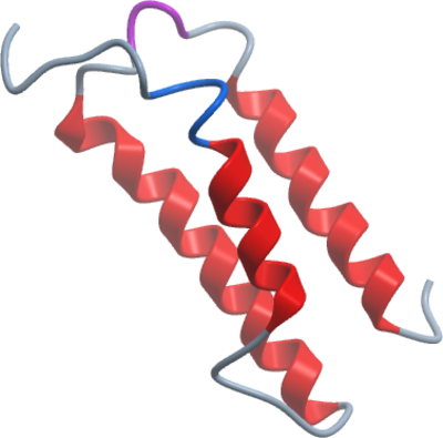
Structure Solution: A total of 12 selenium sites were located (corresponding to six RAMP2 molecules in the crystallographic asymmetric unit) and refined using AUTOSHARP. initial maps allowed the tracing of the entire RAMP2 ECD. Phases were transferred to the native dataset using multi-crystal, six-fold averaging (DMMULTI). Initial model building was carried out using BUCCANEER and completed by hand. |
Resolution: 2.05Å |