SFTPC
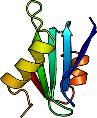
PDB:2YAD
Revision
Revision Type:created
Revised by:created
Revision Date:created
Entry Clone Accession:
Entry Clone Source:FirstChoice PCR-Ready human lung cDNA (Ambion, Cambridgeshire, UK)
SGC Clone Accession:SFTPCA-k013
Tag:N-terminal thioredoxin-tag, hexahistidine tag (integrated thrombin protease cleavage site: LVPRGS) and S-tag (integrated enterokinase cleavage site: DDDK).
Host:E. coli Origami (DE3) pLysS (Novagen)
Construct
Prelude:
Sequence:MSDKIIHLTDDSFDTDVLKADGAILVDFWAEWCGPCKMIAPILDEIADEYQGKLTVAKLNIDQNPGTAPKYGIRGIPTLLLFKNGEVAATKVGALSKGQLKEFLDANLAGSGSGHMHHHHHHSSGLVPRGSGMKETAAAKFERQHMDSPDLGTDDDDKAMAHMSQKHTEMVLEMSIGAPEAQQRLALSEHLVTTATFSIGSTGLVVYDYQQLLIAYKPAPGTCCYIMKIAPESIPSLEALTRKVHNFQMECSLQAKPAVPTSKLGQAEGRDAGSAPSGGDPAFLGMAVSTLCGEVPLYYI
Vector:pET-32c
Growth
Medium:
Antibiotics:
Procedure:SeMet labeled protein
Cells from a glycerol stock were used to inoculate 40 ml LB supplemented with 100 μg/ml ampicillin, and grown at 30 °C overnight. 10 x 15 ml of the overnight culture was used to inoculate 10 bottles with 0.75 l minimal media (without amino acids) supplemented with 8 g/l glycerol, 100 μg/ml ampicillin and approximately 5-10 drops of Dow Corning Antifoam. The culture was grown in a LEX bioreactor system (Harbinger Biotechnology) at 37 °C until OD600 reached ~0.6. The culture was down-tempered to 22 °C over a period of one hour before target expression was induced by addition of 0.5 mM IPTG. Expression was allowed to continue for 9 h. Cells were harvested by centrifugation (4,400 x g, 10 min, 4 °C). The resulting cell pellet (36 g wet cell weight) was resuspended in lysis buffer (2 ml/g cell pellet) supplemented with one tablets of Complete EDTA-free protease inhibitor (Roche Applied Science) and 2000 U Benzonase (Merck) per 100 ml lysis buffer, and stored at -80 °C.
Composition of Minimal media: 25 mM Na2HPO4, 25 mM KH2PO4, 50 mM NH4Cl, 5 mM Na2SO4, 0.4% (w/v) glucose, 2mM MgSO4, 0.1mM CaCl2, 1.0 µM MnCl2, 10 µM FeSO4.
Mix of amino acids added per liter culture (Van Duyne, G. D., J. Mol. Biol. 229, 105-124 (1993)): 100 mg each of lysine, threonine, phenylalanine and 50 mg each of leucine, isoleucine, valine, L(+)-selenomethionine.
Purification
Procedure
Columns:
IMAC: Ni-charged 5 ml HiTrap Chelating HP (GE Healthcare)
IEX: MonoQ 5/50 GL (GE Healthcare)
Gel filtration column: HiLoad 16/60 Superdex 200 Prep Grade (GE Healthcare)
Procedure:
IMAC columns were equilibrated with IMAC wash1 buffer, the IEX column with IEX wash1 buffer, and the gel filtration column with GF buffer. Purification of the protein was performed on an ÄKTA Purifier (GE Healthcare). The filtered lysate was loaded onto the Ni-charged HiTrap Chelating column and washed with IMAC wash1 buffer followed by IMAC wash2 and wash3 buffers. Bound protein was eluted from the IMAC column with IMAC elution buffer. Thioredoxin and hexahistidine tags were released with thrombin at rt and 5 h incubation. The protein was dialyzed against IMAC wash3 buffer and loaded on IMAC columns again. The unbound protein was collected and the tags were eluted with IMAC elution buffer. Fractions containing the target protein were identified by SDS-PAGE, pooled, and loaded to the GF column. Fractions were collected and analyzed by SDS-PAGE. The protein was concentrated using an Amicon Ultra-15 centrifugal filter device (5,000 NMWL; Millipore) to 8-10 mg/m. The identities of the proteins were confirmed by mass spectrometry and selenomethionine was fully incorporated at all methionine positions in the labeled protein. Protein was stored as aliquots at -80 ºC.
Tag removal:
Thrombin was used at enzyme/protein weight ratio of 0.002 percent.
Extraction
Procedure
The cell suspension was quickly thawed in water. Cells were disrupted by sonication (VibraCell, Sonics) at 80% amplitude for 3 min effective time (puls 2s on, 2s off) and centrifuged (49,000 x g, 30 min, 4 ºC). The supernatant was discarded and pellets resuspended in 2 M urea and 20 mM Tris pH 8.0 and incubated at 4 ºC under continuous stirring overnight. The cells were sonicated (VibraCell, Sonics) at 80% amplitude for 3 min effective time (puls 2s on, 2s off) and centrifuged (49,000 x g, 30 min, 4 ºC). The supernatant was stored at 4 ºC.
Concentration:
Ligand
MassSpec:
Crystallization:Crystals were obtained by means of in situ proteolysis with trypsin (1:100 protease/protein ratio) in sitting drop vapour diffusion setups with a 96-well plate. 0.1 µl of the protein solution (10.1 mg/ml) was mixed with 0.2 µl of well solution consisting of 0.1 M bis-Tris pH 6.5, 20% (w/v) PEG MME 5000. The plate was incubated at rt and crystals were obtained after 2 weeks. The crystals were quickly transferred to cryo solution containing well solution and 30% PEG 400 and flash frozen in liquid nitrogen.
NMR Spectroscopy:
Data Collection:Data was collected from one crystal. MAD data was collected to 2.2 Å at ESRF (ID23eh1) Grenoble, France. The crystal belonged to space group C2 with cell parameters of a=132.0 Å b=39.3 Å and c=114.8 Å, and β=99.6.
Data Processing:The structure was solved using SHARP with the three-wavelength MAD data. 18 Se-Met sites were found. Subsequent phase improvement and density modification was done using DM. The structure was rebuilt in Coot and O and refined in Refmac5, Buster and Phenix. Final R-values were R= 19.0% and Rfree= 22.2% and coordinates and structure factors were deposited in the PDB with accession code 2YAD.